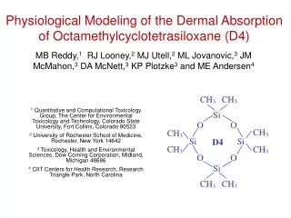
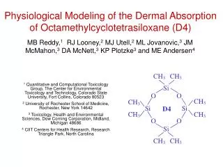
Physiological Modeling of the Dermal Absorption of Octamethylcyclotetrasiloxane (D4)
Physiological Modeling of the Dermal Absorption of Octamethylcyclotetrasiloxane (D4) 1 Quantitative and Computational Toxicology Group, The Center for Environmental Toxicology and Technology, Colorado State University, Fort Collins, Colorado 80523

Physiological Modeling of the Dermal Absorption of Octamethylcyclotetrasiloxane (D4)
E N D
Presentation Transcript
Physiological Modeling of the Dermal Absorption of Octamethylcyclotetrasiloxane (D4) 1 Quantitative and Computational Toxicology Group, The Center for Environmental Toxicology and Technology, Colorado State University, Fort Collins, Colorado 80523 2 University of Rochester School of Medicine, Rochester, New York 14642 3 Toxicology, Health and Environmental Sciences, Dow Corning Corporation, Midland, Michigan 48686 4 CIIT Centers for Health Research, Research Triangle Park, North Carolina MB Reddy,1 RJ Looney,2 MJ Utell,2 ML Jovanovic,3 JM McMahon,3 DA McNett,3 KP Plotzke3 and ME Andersen4
Abstract • Studies of human dermal absorption of octamethylcyclotetrasiloxane, D4, through axilla skin in vivo and through abdominal skin in vitro have recently been completed. A mathematical model describing the dermal absorption of D4 was developed and combined with an inhalation PBPK model for this material. The model includes volatilization of D4 from the skin surface, evaporation of chemical out of the skin after the skin surface had been cleared of the chemical, and a deep skin compartment. The in vivo dermal absorption study of D4 in the rat provided evidence that a model structure including elimination from the skin by evaporation was appropriate. Concentrations of D4 in exhaled air and blood plasma from human, in vivo exposures were used to estimate the model parameters. Following either inhalation or dermal exposures, D4 blood plasma concentrations increased with time relative to exhaled air concentrations. The PBPK model for both dermal and inhalation exposures required the inclusion of a pool of unavailable D4 created in the liver, transported in the blood, and cleared in the liver to describe this behavior. Model calculations indicated that during the human, in vivo, dermal exposure, more than 90% of the applied dose evaporated from the skin surface before it could be absorbed into the skin. Of the D4 absorbed into the skin, the majority was eliminated by evaporation before systemic absorption could occur. For men and women, respectively, about 0.1 and 0.5% of the applied dose of D4 entered the cutaneous blood within 24 hours of the exposure.
Introduction • Octamethylcyclotetrasiloxane (D4) is a lipophilic (logKo/w 5), semi-volatile (vapor pressure 0.68 mmHg at 20C) compound primarily used as an intermediate in the manufacturing of high molecular weight silicone polymers and as an ingredient in some consumer products. • Recently, several studies evaluating the dermal absorption of D4 have been completed: • in vivo dermal absorption through human axilla skin • in vitro dermal absorption through human abdomen skin • in vivo dermal absorption through rat skin • Here, we analyze and interpret these data. • A compartment model describing the dermal absorption of volatile chemicals was combined with a human D4 PBPK model for the analysis of the human, in vivo, dermal absorption data.
Background • During dermal absorption, the absorbingchemical must diffuse through the stratum corneum and viable epidermis. Once the chemical reaches the highly vascularized dermis, it enters the blood stream and systemic circulation. For clarity, we define the following: The amount absorbed is the amount ofchemical that has passed through the skin into the blood combined with the amount of chemical remaining in the viable skin layers. The amount penetrated is the amount of chemical that has passed through the skin into the blood. • Often, the amount absorbed (particularly in vitro) is used as an estimate of the amount penetrated for lipophilic materials
Model • For dermal exposures to neat, volatile or semi-volatile chemicals, the skin surface is exposed to the chemical until all of the chemical has left the skin surface due to evaporation or dermal absorption. • While D4 remains on the skin, the mass transfer of D4 from the skin surface by evaporation was modeled as a zero-order process and the dermal absorption of D4 was modeled as a first-order process (Figure 1). • After the skin cleared of chemical (i.e., after all the D4 left the skin surface by evaporation or dermal absorption), the model included D4 mass transfer from the skin back into the air. • The model also included a deep skin compartment. • The dermal absorption model was combined with a human inhalation PBPK model for D4 (Figure 1, Table 2), which required several features for describing D4 kinetics: • mass transfer limitations in the slowly perfused compartment and fat tissue • a pool of unavailable D4 that was produced in the liver, traveled through the bloodstream, and cleared in the fat • PBPK model equations were solved using Berkeley Madonna and the multiple curve-fitting algorithm was used to fit the model output to human, in vivo data (i.e., Experiment 1 data) to calculate parameters for the dermal absorption model (Table 1).
Table 1. Parameters for the dermal absorption model. men 0.0098 0.00050 3.8 0.014 0.060 0.010 women 0.0068 0.00015 0.18 0.036 0.038 0.000010 kv, g/cm2/min k1, cm3/min k-1, min-1 k-2 , min-1 kd , min-1 k-d , min-1
Cin alveolar space Cout lung blood (a) exposed skin venous blood arterial blood slowly perf. tissue rapidly perf. tissue fat deep fat blood lipid liver metabolism (continued)
(continued from last page) (b) venous blood clearance in fat venous blood productionin liver (c) evaporation of D4 onthe skin surface evaporation of D4 that has absorbed into the skin kv neat D4 k-1 k-2 venous blood skin k1 k-2 venous blood skin kd k-d deep comp. kd k-d deep comp. Figure 1. Schematic diagram of (a) the PBPK model [1, 4], (b) the sub-model for the pool of unavailable D4, and (c) the compartment model of dermal absorption before and after the D4 dose has evaporated or absorbed into the skin.
Table 2. Parameters used in the D4 human PBPK model [4]. Parameter QP QC liver fat rapidly perf. tissue slowly perf. tissue liver fat rapidly perf. tissue slowly perf. tissue Value 7.6 L/min 5.9 L/min 0.227 0.052 0.472 0.249 0.0314 0.23 0.05 0.5396 alveolar ventilation cardiac output fraction of blood flow to tissues fraction of body weight in tissues (continued)
(continued from last page) allometric constant formetabolic clearance allometric constant forvolume of distribution clearance for metabolites blood:air liver:blood fat:blood slowly perf. tissue:blood rapidly perf. tissue:blood slowly perfused comp. mass transfer into deep fat mass transfer from deep fat first order production rate clearance into fat 0.097 L/min/kg0.7 1.2 L/kg 0.038 L/min 0.94 8.9 490 3 8.4 0.36 L/min 0.0038 min-1 0.0021 min-1 0.053 min-1 0.014 L/min parameters for metabolism partition coefficients parameters for mass transfer limitations in tissues production and clearance of unavailable D4 in blood
Experiment 1Dermal Absorption through Human Skin In Vivo • Three male and three female subjects had 0.7 and 0.5 g of [13C]-D4 applied to each axilla, respectively. For all subjects, there was a 5-min pause before the test chemical was applied to the second axilla. This work was conducted at the University of Rochester. • After the exposure, samples of expired air were collected in a 5-liter Tedlar bag using a Hans Rudolf non-rebreathing valve . • The amount of [13C]-D4 in plasma and exhaled air (Figure 2) was measured using GC/MS. Figure 2. Measured (symbols) and calculated (solid lines) D4 concentrations in (A) exhaled breath and (B) blood plasma for men () and women () after a dermal exposure. The error bars represent one standard deviation for n = 3.
Experiment 2Dermal Absorption through Rat Skin In Vivo • A 2.5 cm2 aluminum skin depot was glued to the back of F344 female rats housed in Roth-style glass metabolism cages for the collection of expired air and excreta. • For all three dose levels, 1.9, 4.8 and 9.7 mg/cm2, groups of 4 rats were sacrificed at 1, 6 and 24 h following the exposure. Another group of rats was washed at 24 h but notsacrificed until 168 h. • At the time of sacrifice, the exposed site was wiped, washed with a soap solution, dried, washed with 70% ethanol, dried, and then tape stripped to remove the stratum corneum. • The amount of 14C in the urine, feces, skin depot, charcoal basket, skin washes, tapes, excised skin at the exposure site, carcasses, the CO2 and volatiles absorbents, and cage washes was determined by liquid scintillation counting. • The amount absorbed was calculated as the amount expired either as parent compound or CO2 and the amount in the urine, feces, excised skin, carcass. The amount penetrated included the same sans D4 in excised skin (Figure 3).
Figure 3. For Experiment 2, the cumulative amount of D4 absorbed and penetrated as a function of time for all doses. The skin of rats sacrificed at 168 h was cleaned 24 hours following the exposure. Points are connected; the lines do not represent model simulation.
Experiment 3Dermal Absorption through Human Skin In Vitro • Skin disks obtained from abdominal skin of 6 human cadavers were dermatomed to a thickness of 356-457 m and mounted on Bronaugh flow-through diffusion cells in a 32C water bath. • Skin integrity was verified using [3H]-H2O. • About 10.7 mg/cm2 [14C]-D4 was applied to an exposure area of 0.64 cm2 on the skin. The application site was covered with a charcoal basket to trap any D4 that evaporated. • The receptor medium (Hank’s Balanced Salt Solution with 0.6% HEPES, 0.005% Genetecin and 4% BSA adjusted to a pH of 7.4) flowed continuously through the receptor chamber and was collected directly into liquid scintillation vials for the determination of the cumulative amount of D4 penetrated (Figure 4).
applied dose of D4: - 11 mg/cm2 - 16 mg/cm2 - 7.3 mg/cm2 - 8.4 mg/cm2 - 13 mg/cm2 - 7.9 mg/cm2 Figure 4. For Experiment 3, the cumulative amount of D4 that penetrated through human skin in vitro (i.e., into receptor medium) as a function of time for six experiments. Points are connected; the lines do not represent model simulation.
Results: Model Structure • The model for describing the dermal absorption of D4 included volatilization of neat D4 from the skin surface, evaporation of D4 from the skin after the skin had cleared of chemical, and a deep skin compartment (Figure 1c). • Dermal absorption models do not usually include evaporation of chemical back out of the skin following an exposure, but the in vivo dermal absorption study of D4 in the rat (Figure 3) provided evidence that this model structure was appropriate. During Experiment 2: • The amount of D4 that absorbed decreased significantly with time, but there were no corresponding increases in the amount penetrated. This provides evidence that some D4 was eliminated from the skin by evaporation before penetration occurred. • For volatile or semi-volatile chemicals that can be eliminated from the skin by evaporation, the amount absorbed may be significantly higher than the amount penetrated. • Calculated D4 plasma concentrations more closely matched the experimental data than calculated concentrations in exhaled air because the human inhalation PBPK model also matched blood plasma concentration data more closely. • Peak D4 plasma and exhaled air concentrations probably occurred before the earliest samples were collected at one hour following the exposure. Because data were unavailable at early times when peak blood concentrations and D4 evaporation occurred, model predictions at early times require confirmation. • Model parameters were calculated for men and women separately (Table 1) because D4 concentrations in plasma and exhaled air were higher for women than for men.
Results: Calculations for Experiment 1 • By including the dermal exposure route in a human D4 inhalation PBPK model, it was possible to calculate that during human, in vivo, dermal exposure (Experiment 1): • all the applied D4 would be cleared from the skin within 5 minutes due to evaporation of neat D4 and dermal absorption • more than 90% of the applied dose evaporated from the skin surface before it could be absorbed into the skin • the majority of the D4 that had absorbed into the skin was eliminated from the skin by evaporation before penetration into systemic blood could occur • the maximum D4 plasma concentration was more than 100 mg/L • for men and women, respectively, about 0.1 and 0.5% of the applied dose of D4 penetrated the axilla skin in 24 h following a dermal exposure • The calculation that more than 90% of the applied neat D4 evaporated is consistent with the other experiments: • Experiment 2: more than 92% of the applied dose was recovered from the charcoal filter covering the exposed site at 24 hours for all doses • Experiment 3: an average of 88.2% of the applied dose was recovered from the charcoal filter covering the exposed site at 24 hours • For Experiment 1, with an average applied dose of 30.5 mg/cm2, about 0.3% of the applied dose penetrated. For Experiment 3, with an average applied dose of 10.7 mg/cm2, about 0.01% of the applied dose penetrated. This discrepancy could be because of different doses or regional differences in skin properties (e.g., axilla skin has been shown to absorb more parathion, malathion and hydrocortisone than other regions [2,3]). • For Experiment 1, the amount penetrated could be calculated because the data contained information pertaining to the amount of D4 that reached systemic circulation (e.g., plasma concentrations), but the amount absorbed could not be calculated.
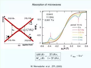
Model Structure • After an inhalation exposure has ended and during dermal exposures, the ratio of the concentration of chemical in the venous return to the concentration in exhaled breath (i.e., Cv/Cex) is expected to remain constant over time [4]. • Surprisingly, after human inhalation exposures to 10 ppm [14C]-D4, the ratio Cv/Cex increased with time (Figure 5). To describe this behavior, the PBPK model was modified to include a pool of unavailable D4 that was produced in the liver, moved through the blood, and was cleared in the fat (Figure 1) [1]. • Because the ratio Cv/Cex also increased with time following the human, in vivo, dermal exposures, the same model structure is appropriate for inhalation and dermal exposures and blood sequestration of D4 is equally important for describing D4 kinetics following dermal and inhalation exposures. Figure 5. The ratio Cv/Cex as a function of time for inhalation () and dermal () exposures.
Summary • The dermal absorption model included volatilization of D4 from the skin surface, evaporation of D4 out of the skin after the skin surface had been cleared of the chemical, and a deep skin compartment. • By including the dermal exposure route in a human D4 inhalation PBPK model, it was possible to calculate that during human, in vivo, dermal exposure (Experiment 1): • more than 90% of the applied dose evaporated from the skin surface before it was absorbed • of the D4 that was absorbed into the skin, most was eliminated by evaporation before penetration into systemic blood occurred • about 0.3% of the applied dose of D4 penetrated in 24 hours • For highly lipophilic and semi-volatile chemicals that can eliminate from the skin by evaporation, the amount penetrated may be significantly less than the amount absorbed.
References [1] Andersen, ME, Sarangapani, R, Reitz, RH, Gallavan, RH, Dobrev, ID and Plotzke, KP. 2001. Physiological modeling reveals novel pharmacokinetic behavior for inhaled octamethylcyclotetrasiloxane in rats. Toxicol Sci60:214-231. [2] Feldmann, RJ and Maibach, HI. 1967. Regional variation in percutaneous penetration of 14C cortisol in man. J Invest Dermatol48:181-183. [3] Maibach, HI, Feldmann, RJ, Milby, TH, and Serat, WF. 1971. Regional variation in percutaneous penetration in man - pesticides. Arch Environ Health23:208-211. [4] Reddy, MB, Andersen, ME, Morrow, PE, Dobrev, ID, Varaprath, S, Plotzke, KP and Utell, MJ. In press, 2003. Physiological modeling of inhalation kinetics of octamethylcyclotetrasiloxane (D4) in humans during rest and exercise.Toxicol Sci. Acknowledgements • M. Reddy received support from grant number F32 ES11425-02 from the National Institute of Environmental Health Sciences (NIEHS), NIH. This work is solely the responsibility of the authors and does not necessarily represent the official views of NIEHS, NIH. The support of many of our colleagues at CETT, especially that of R. Yang, is gratefully acknowledged.