Download
1 / 41
410 likes | 644 Views
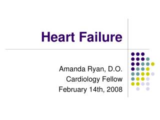
C ONGENITAL HEART DİSEASES. DR DEFNE ÇÖL YEDİTEPE UNIVERSITY DEPARTMENT OF CHİLD HEALTH AND DİSEASES. Fetal Circulation.

E N D
CONGENITAL HEART DİSEASES DR DEFNE ÇÖL YEDİTEPE UNIVERSITY DEPARTMENT OF CHİLD HEALTH AND DİSEASES.
FetalCirculation Fetal circulation differs from adult circulation in several ways. Almost all differences are attributable to the fundamental difference in the site of gas exchange. In the adult, gas exchange occurs in the lungs. In the fetus, the placenta provides the exchange of gases and nutrients.
CONGENİTAL HEART DİSEASE(CHD) • Congenital • Anatomic>abnormal function • Acquired • Disease process • Infection • Autoimmune response • Environmental factors • Familial tendencies
Chromosomal/genetic = 10%-12% • Maternal or environmental = 1%-2% • Maternal drug use • Fetal alcohol syndrome—50% have CHD • Maternal illness • Rubella in 1st 7 wks of pregnancy→50% risk of defects including PDA and pulmonary branch stenosis • CMV, toxoplasmosis, other viral illnesses>> cardiac defects • IDMs = 10% risk of CHD (VSD, cardiomyopathy, TGA most common) • Multifactorial = 85%
Incidence: 5-8 per 1000 live births • About 2-3 of these are symptomatic in first year of life • Major cause of death in first year of life (after prematurity) • Most common anomaly is VSD • 28% of kids with CHD have another recognized anomaly (trisomy 21, 13, 18, +++ )
Alagillesnydrome Williams snydrome Trisomi 18 Trisomi 13
Pediatric Indicators of Cardiac Dysfunction • Poor feeding • Tachypnea/ tachycardia • Failure to thrive/poor weight gain/activity intolerance • Developmental delays • + Prenatal history • + Family history of cardiac disease
Older Classifications of CHD • Acyanotic • May become cyanotic • Cyanotic • May be pink • May develop CHF
Newer Classification of CHD • Hemodynamic characteristics • Increased pulmonary blood flow • Decreased pulmonary blood flow • Obstruction of blood flow out of the heart • Mixed blood flow
Increased Pulmonary Blood Flow Defects • Atrialseptal defect • Ventricular septal defect • Patent ductusarteriosus
AtrialSeptalDefect • İncidence: 5-10% • Pathology:Classifiedaccording to location of defect: • Secundumatrialseptaldefect: • Sinusvenosusatrialdefect: • Primumatrialseptaldefect:
ClinicalManifestations • Small and moderate size atrialseptal defects are typically asymptomatic. Largerdefects result in pulmonary edema manifesting as easy fatigability and shortness ofbreath. Only very large defects result in significant congestive heart failure. • On examination:there is a hyperactive precordium with a prominent rightventricularimpulseduetorightventriculardilation. Auscultationreveals a prominentfirst heart sound. Second heart splitting is fixed throughout respiration due toincreased blood flow through the pulmonary valve causing delay in pulmonaryvalve closure regardless of respiratory cycle. A systolic ejection (crescen-decrescendo) murmur is heard at the left upper sternal border due to increase inblood flow across the pulmonary valve.
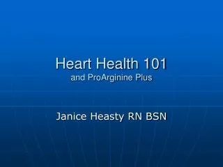
Diagnosis X-Ray:Prominent pulmonary vasculature due to left to right shunting is present.Inaddition, increase in blood flow through the right heart will cause right atrialand right ventricular dilation manifesting as cardiomegaly on chest X-ray. Electrocardiograph:Right atrial and right ventricular dilation/hypertrophy may be noted. Right atrialenlargementmanifests as tall P waves Echocardiography:The atrialseptal defect is seen by 2D echocardiography.The right atrium and ventricle will appear dilated.
EKG X_Ray
Treatment • Closure of atrialseptal defect is determined by the type of the defect and itssize. Small (less than 5 mm in diameter) and medium (5–8 mm in diameter)-sizedsecundumdefects diagnosed during early infancy tend to close spontaneously,often in the first 2 years of life. • Sinusvenosusandprimumatrialseptaldefects do not close spontaneously and will require surgical repair which could be performedaround 1 year of age.
VentricularSeptalDefect • İncidence:20-25% Ventricular septaldefect is themost common cardiac defect. Membranous ventricular septal defect is the most common type (70%). Theefectoccurs in the membranous septum andinvolves some of the surrounding tissue. Inlet (AV canal type) ventricular septal defect accounts for 5–8% of all ventricularseptaldefects. Muscular ventricular septal defect accounts for 5–20% of all ventricular septaldefects. It is located in the muscular septum. Outlet (infundibular, conal, and supracristal) ventricular septal defectaccount for 5–7% of all types of defects. The defect is locatedintheoutlet septum, beneathboth semilunar (pulmonary and aortic) valves.
Pathophysiology • In small ventricular septal defects the defect is restrictive and the amount ofshunting will be hemodynamically insignificant. If the defect is large there will besignificant shunting to the right side depending primarily on the difference betweenthe systemic and pulmonary vascular resistance , this will cause dilatation. of the pulmonary arteries, left atrium, and left ventricle. The excessive shuntingwill also cause increase in pulmonary blood flow and congestive heart failure secondarytovolumeoverload.
ClinicalManifestations:Most infants with small ventricular septal defects are asymptomatic. Presentation istypically secondary to a heart murmur on physical examination. In moderate to large ventricularseptaldefect, the infants present with symptoms secondary to increased pulmonary blood flow (pulmonary edema) and decrease in cardiac output such as tachypnea,increased respiratory effort, recurrent pulmonary infections, poor feeding, diaphoresis,easyfatigability, and failure to thrive. Older patients may present with heartfailure, hemoptysis, arrhythmia, cyanosis, orbacterialendocarditis. Ventricular septal defect murmurs may be 2–5/6 in intensity and harsh in quality,itis best heard over the left lower sternalborder.
ChestRadiography:The chest X-ray is normal in small ventricular septal defects. In moderate and largeventricular septal defects there is usually cardiac enlargement withincreasedpulmonaryvascularmarkings. Electrocardiography:Leftatrial dilatation and left ventricular hypertrophy may be seen in moderate ventricularseptal defect. With a large defect, the ECG shows biventricular hypertrophy. Echocardiography:The echocardiogram is the gold standard tool to diagnose ventricular septaldefect.It can identify the size, location, and number of ventricular septal defects.
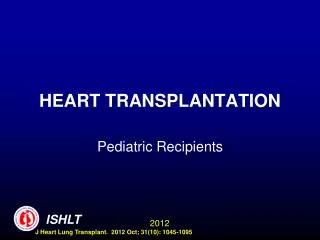
a 3-month-old infant with a large ventricular septal defect, patent ductus arteriosus, and pulmonary hypertension. The tracing shows combined ventricular hypertrophy with left dominance. Note that V2 and V4 are in ½ standardization.
Patent DuctusArteriosus The incidence of PDA isinversely related togestational age in prematureinfants(5-10%) In normal newborns, the ductus is mostly closed by thesecond or third day of life and is fully sealed by 2–3 weeks of life. With the baby’s first few breaths, the oxygentensionrises. the pulmonary vascular resistance begins to drop. If the ductusarteriosusfails to close, there will be shunting of blood from the high pressure aorta to thepulmonarycirculation.
ClinicalManifestations If the PDA is small, patients are typicallyasymptomatic. A large PDA will allow asignificant volume of left to right shunting. The resulting pulmonary edema canmanifest clinically as tachypnea, poor feeding, failure to thrive, recurrent respiratoryinfections, or congestive heart failure. • Murmurof a PDA is acontinuous machinery murmur and is heard best in the left infraclavicularregion.
Electrocardiography: Electrocardiogram is usually normal with a small PDA. Left atrial and ventricularhypertrophy may be present in older patients with a moderate sized PDA Biventricular hypertrophy is present in patients with a large PDA. Echocardiography: Echocardiography is the procedure of choice to confirm the diagnosis Managementindomethacinandibuprofenhave been used for their antagonizing effects on prostaglandins. interventionalcardiac catheterization
Obstructive Defects • Coarctation of the aorta • Aortic stenosis • Pulmonicstenosis
İncidence:8-10% ,M/F : 2/1 • Higher blood pressure in upper extremitieswhen compared to blood pressurein lower extremities is diagnostic of coarctation of the aorta. • Increased afterload results inleftventricularhypertrophy. • Coarctation of the aorta may present inchildhood or adulthood with systemichypertension, usually resistant to medications.
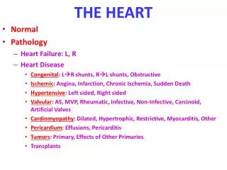
Rib notching (arrows) in an 11-year-old girl with coarctation of the aorta. The figure-of-3 configuration indicates the site of coarctation with the large proximal segment of aorta and/or prominent left subclavian artery above and the poststenotic dilatation of the descending aorta below it. B, Barium esophagogram reveals the E-shaped indentation or reversed figure-of-3 configuration
AorticStenosis Anatomictypes of aorticstenosis. A, Normal. B, Valvularstenosis. C, Supravalvularstenosis. D, Discretesubaorticstenosis. E, Idiopathichypertrophicsubaorticstenosis Occurring in approximately 10% of cases of congenital heart disease, aortic stenosis refers to obstruction to outflow from the left ventricle due to narrowing at above, below, or at the level of the aortic valve. Narrowing at the aortic valve (valvular aortic stenosis) accounts for 71% of cases of aortic stenosis.
ClinicalManifestations: Theleftventriclegraduallyhypertrophies in order to accommodate the increased force necessary for aorticvalve opening. As hypertrophy eventually gives way to left ventricular failure, theleft ventricle and left atrium dilate and changes related to increased left ventricularend-diastolic pressure and left atrial hypertension occur.Patientspresent with symptoms of syncope, chest pain, anddyspnea, typicallywithexertion. Newborn children with critical aortic stenosis present in shock-likestate within the first hours to 1 month of life as ductal closure leads to reducedantegradeflow blood flow across the aortic valve.
PulmonaryStenosis As an isolated lesion, valvular pulmonary stenosis is the second most commonCHD (8% of all CHDs). Pulmonary stenosis at some level,whethervalvular(90%),subvalvular,orsupravalvular, occurs in 30–50% of othercongenital heart diseases. Pulmonary stenosis occurs morefrequently in females. Supravalvularpulmonary stenosis also occurs as a result ofintrauterine (congenital)rubellainfection.
ClinicalManifestations: right ventricle is hypertrophie. Moderate to severe pulmonary artery stenosis may result in fatigue andreduced exercise tolerance. Moderate valvularstenosis is often well toleratedin children, but produces clinical symptoms with advancing age. Severevalvularstenosis can lead to exercise-related chest pain, syncope, or suddendeath.
Decreased Pulmonary Blood Flow Defects • Tetralogy of Fallot. • PulmonaryAtresiawithIntactVentricularSeptum. • TricuspidAtresia.
Tetralogy of Fallot Tetralogyof Fallot is themost common cyanotic congenital heartdisease(10%) As the name implies, there are four basic components that make up TOF : • 1. A largeventricularseptaldefect (VSD) • 2. Pulmonarystenosis (PS) • 3. An overriding aorta • 4. Right ventricular hypertrophy (RVH)
TOF is well tolerated in utero. Once born, newborn children are frequentlyasymptomaticand often do not exhibit cyanosis. Infants and children with unrepaired TOF are at risk for episodes of severecyanosis known as hypercyanoticspells, commonly referred to as “tet spells.” These episodes rarely occur in children less than 9–12 months of age In the hospital setting, treatment of hypercyanotic spells should start withattempts to reduce any cause of anxiety to the child. Allow the child’s mother tohold him or her in a knee-to-chest position to increase systemic vascular resistance.
ChestX-Ray:The apex can seem to be upturned due to RVHresulting in the classically described “coeur en sabot” or boot-shaped heart. • Treatment:In the modern era of congenital heart surgery, with patients being successfully operatedon at smaller weights and younger ages with excellent results, it is now oftenpossible for patients to undergo complete anatomic repair as their initial operation. Complete repair ofTOF can be safely performed at 4–6 months of age.
Transposition of the Great Arteries Transposition of the great arteries is a cyanotic congenital heart diseases where thegreat arteries (pulmonary artery and aorta) are connected to the wrong ventricle.
Total Anomalous Pulmonary Venous Return Total anomalous pulmonary venous return (TAPVR) is a cyanotic congenital heartdisease where blood from all four pulmonary veins returns anomalously to the rightatrium instead of the left atrium.
TruncusArteriosus Truncusarteriosus is a cyanotic congenital heart disease. In this lesion, there is onlyone (truncus) artery receiving blood ejected from both ventricles. The truncusthencontinue as the aortic arch and providing pulmonary arteries.
Ebstein’sAnomaly Ebstein’sanomaly is a congenital heart disease affecting the tricuspid valve. In itsmilder form, the tricuspid valve is mildly displaced towards the apex with mildregurgitationand no stenosis. tricuspid valve leaflets results in severetricuspid valve regurgitation and lack of forward flow of blood in the right ventricularoutflowtract due to obstruction by the abnormal tricuspid valve.
HypoplasticLeftHeartSyndrome Themitral valve is severely stenotic or atretic leading to small or hypoplastic left ventricleand severely stenotic or hypoplastic aortic valve. Neonates survive this anomaly in the first few days of life dueto the presence of a patent foramen ovale (PFO) and ductusarteriosus (PDA) Cardiogenicshock develops as soon as the PDA starts to close depriving cardiac output to thesystemiccirculation.