Scaffold-Free Fabrication of Tubular Cellular Constructs via Inkjet Printing
This study explores the innovative approach of scaffold-free organ printing using piezoelectric-driven inkjet technology to create tubular cellular constructs. By leveraging a bioink composed of 3T3 fibroblast cells and sodium alginate, the research successfully fabricated three-dimensional constructs with consistent diameters of 2-3 mm and a wall thickness of approximately 100 µm. Notably, cell viability post-printing exceeded 80%, demonstrating the feasibility of maintaining cellular functionality within the printed constructs, thus paving the way for future advancements in organ transplantation.

Scaffold-Free Fabrication of Tubular Cellular Constructs via Inkjet Printing
E N D
Presentation Transcript
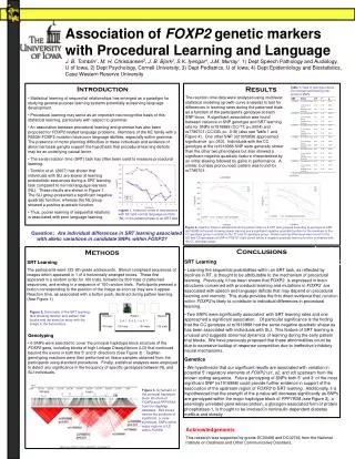
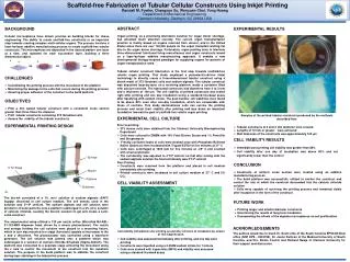
D C B A Scaffold-free Fabrication of Tubular Cellular Constructs Using Inkjet Printing Randall M. Fowler, Changxue Xu, Wenxuan Chai, Yong Huang Department of Mechanical Engineering Clemson University, Clemson, SC 29634 USA ABSTRACT Organ printing, as a promising alternative solution for organ donor shortage, has attracted much attention recently. The current organ transplantation practice is mainly based on organs received from donors; and in the United States alone there are over 100,000 people on the organ transplant waiting list due to the organ donor shortage. Fortunately, organ printing aims to fabricate three-dimensional functional living macrotissues and organ constructs based on a layer-by-layer additive manufacturing approach. It enables a new developmental biology-inspired paradigm for supplying organs for patients of organ transplantation need. Tubular cellular construct fabrication is the first step towards scaffold-free robotic organ printing. This study employed a piezoelectric-driven inkjet technology to directly create a three-dimensional tubular construct using a bioink made of 3T3 fibroblast cells and sodium alginate. The tubular construct was deposited layer-by-layer on a receiving platform inside a container filled with calcium chloride. The fabricated constructs had diameters from 2 to 3 mm and a thickness of 100 um. The cell viability of printed constructs was tested right after printing and one day incubation using a standard live-dead assay after liquefying with sodium citrate. The post-transfer cell viabilities were found to be above 80% even after one-day incubation, which are comparable with those of controls. This study demonstrates cells can survive the printing process and retain their viability after printing and lays down an important foundation towards the goal of scaffold-free robotic organ printing. EXPERIMENTAL RESULTS BACKGROUND Cellular microspheres have shown promise as building blocks for tissue engineering. The ability to create scaffold-free constructs is an important step towards creating complex multi-cellular organs. The process involves a layer-by-layer additive manufacturing process to create scaffold-free tubular constructs. The microspheres are deposited in the desired pattern one layer at a time, and repeated for each successive layer, building a three dimensional object. • CHALLENGES • Coordinating the printing process with the movement of the platform • Minimizing the damage to the cells that occurs during the printing process • Assuring proper adhesion of the construct to the build platform • OBJECTIVES • Print a thin walled tubular construct with a consistent cross section greater than three times the diameter • Print tubular constructs containing 3T3 fibroblast cells • Assess the viability of the tubular constructs Samples of the printed tubular constructs produced by the methods described here EXPERIMENTAL CELL CULTURE EXPERIMENTAL PRINTING DESIGN • Prior to printing • 3T3 mouse cells were obtained from the Clemson University Bioengineering Department • Cells were cultured in DMEM with 10% Fetal Bovine Serum and 1% Penicillin and Streptomycin • Freshly confluent flasks of cells were washed once with Phosphate Buffered Saline (Dubecco) then incubated with Trypsin/EDTA for five minutes at 37° C • Cells were centrifuged at 1000 rpm for five minutes at ~25° C and counted with a hemocytometer • The cell density was adjusted to 4*106 cells/ml so that after mixing with the sodium alginate solution the final cell density was 2*106 cells/ml • Post Printing • Constructs were removed from the platform and placed in cell medium immediately after printing • Printed constructs were incubated in cell culture medium at 37° C and 5% CO2 • Tubular constructs of 2 and 3 mm diameter were created • Lengths of 10 mm or greater were achieved • Wall thickness of the constructs was approximately 100 µm CELL VIABILITY RESULTS • Immediate post printing cell viability was greater than 80% • Cell viability after one day of incubation was above 80% and not significantly lower than the control Syringe • CONCLUSION • Constructs of uniform cross section were created using an additive manufacturing process • The build platform was successfully utilized to anchor the construct and control the rate at which the construct descended into the calcium chloride solution • Cells were capable of surviving the printing process and remained viable after incubation in the form of the construct X YZ Stage Nozzle Platform CELL VIABILITY ASSESSMENT Substrate a b The bio-ink consisted of a 1% (w/v) solution of sodium alginate (SAFC Supply) dissolved in cell culture medium. The cell density used in the solution was 2*106 cells/ml. The sodium alginate and cell solution were printed in circular patterns onto a platform submerged in a 2% (w/v) solution of calcium chloride, causing the bio-ink solution to gel and create a semi-solid construct. The experimental setup utilized a 120 µm nozzle orifice (MicroFab MJ-ABL-01-120-6MX dispense head, driven by a sleeve piezoactuator). The nozzle and syringe holding the cell solution were placed in a mounting fixture, which in turn was mounted to a stage (Aerotech) capable of movement in the x and ydirections. The piezoactuator was controlled using a waveform generator. The cell solution was printed onto a platform which was submerged in a solution of calcium chloride dihydrate (Sigma-Aldrich). The platform was connected to a separate stage achieving the movement along the z axis to control the movement of the construct into the substrate solution. The purpose of the build platform was to stabilize the construct during layer stacking in the fabrication process. FUTURE WORK • Printing longer and smaller diameter constructs • Determining the results of long term incubation • Documenting the effects of the alginate microspheres on cell proliferation ACKNOWLEDGEMENTS The authors would like to thank Dr. Scott Little of the South Carolina EPSCoR/IDeA office (NSF EPS - 0903795), Dr. Joann Sullivan of the Medical University of South Carolina, and Drs. Nicole Coutris and Richard Swaja of Clemson University for their support and discussion. Cell viability immediately after printing (a) and after 24 hours of incubation (b), shown at 10X magnification. • Cell viability was assessed immediately after printing, and oneday post printing • Constructs were liquefied using a 0.055M sodium citrate for 1 minute • Cells were stained with trypan blue (MFG) and viability was assessed using a standard live/dead assay