Download
1 / 72
720 likes | 910 Views
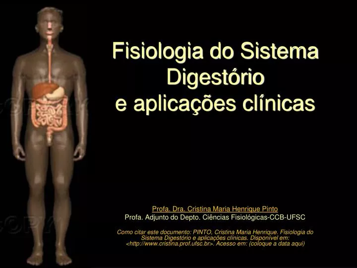
Fisiologia do Sistema Digestório e aplicações clínicas. Profa. Dra. Cristina Maria Henrique Pinto Profa. Adjunto do Depto. Ciências Fisiológicas-CCB-UFSC

E N D
Fisiologia do Sistema Digestório e aplicações clínicas Profa. Dra. Cristina Maria Henrique Pinto Profa. Adjunto do Depto. Ciências Fisiológicas-CCB-UFSC Como citar este documento: PINTO, Cristina Maria Henrique. Fisiologia do Sistema Digestório e aplicações clínicas. Disponível em: <http://www.cristina.prof.ufsc.br>. Acesso em: (coloque a data aqui)
Esta apresentação foi utilizada em minhas aulas para a graduação em Medicina (6ª fase) até o ano de 2007. Para que este material não se perca, deixo aqui à disposição daqueles que eventualmente tenham interesse. Bons estudos!
Bibliografia básica recomendada sobre Fisiologia Humana Livros-textos: “Berne & Levy: Fisiologia” Koeppen & Stanton, 2009, 6ª Ed. (Ed. Elsevier) “Tratado de Fisiologia Médica” Guyton & Hall, 2006, 11ª Ed. (Ed. Elsevier) “Fisiologia” Aires, M. M. 2008, 3ª Ed. (Ed. Guanabara Koogan) “Fisiologia” Costanzo, 2007, 3ª Ed. (Ed. Elsevier) “Berne & Levy: Fundamentos de Fisiologia”, Levy et al, 2006, 4ª Ed. (Ed. Elsevier) “Fundamentos de Fisiologia Médica” Johnson, 2003 (Ed. Guanabara Koogan) “Fisiologia: texto e atlas” Silbernagl e Despopoulos, 2003 (Ed. Artmed)
O Pâncreas como glândula exócrina Profa. Dra. Cristina Maria Henrique Pinto CFS/CCB – 6ª fase - Medicina
PÂNCREAS localização: -órgão retroperitoneal. -anteriores a ele estão o estômago, alças do intestino delgado, colon e omento divisões: cabeça e processo uncinado, colo, corpo e cauda Figure 3. The pancreas and adjacent anatomy. http://hopkins-gi.nts.jhu.edu/pages/latin/templates/index.cfm?pg=disease1&organ=4&disease=22&lang_id=1
Ducto pancreático principal formado a partir dos ductos intercalares, intralobares extralobares e ductos coletores principais Duct system of the pancreas. Berne et al., 2004
Ducto pancreático: -desde a cauda até a cabeça do pâncreas -une-se ao ducto colédoco já próximo ao duodeno, formando a ampola hepatopancreática e a papila maior do duodeno (ou ampola de Vater). É comum a existência de um ducto pancreático secundário, acessório ou dorsal (papila menor) http://hopkins-gi.nts.jhu.edu/pages/latin/templates/index.cfm?pg=disease1&organ=4&disease=22&lang_id=1
DUCTO PANCREÁTICO PRINCIPAL E DUCTO PANCREÁTICO ACESSÓRIO (ocorrência em 90% da população) Normally (90% of the time) pancreatic secretions from the entire pancreas and biliary secretions gain access to the duodenum by way of the ventral pancreatic or Wirsung’s duct because of the fusion of the dorsal pancreatic duct during embryological development. There is only a remnant of the dorsal pancreatic or Santorini’s duct connecting to the duodenum. In 10% of individuals, fusion does not occur. This situation is called pancreas divisum. In pancreas divisum secretions from the dorsal and ventral ducts drain separately into the duodenum. Individuals with pancreas divisum may be at higher risk for pancreatitis (see Acute Pancreatitis for additional information). Note that the distal common bile duct reaches the duodenum through the head of the pancreas. http://www.gastroslides.org/main/browse_deck.asp?tpc=2&mxpg=390&pg=1844#image
EMBRIOLOGIA This figure demonstrates that the stomach, duodenum, liver, biliary system, gallbladder and pancreas are derived from closely related structures in early embryological development. The pancreas, liver, gallbladder and biliary system bud from the duodenum during early embryological development. The pancreas starts as two components, the ventral and dorsal pancreas. In the process of development, the organs enlarge and the ventral pancreas together with the common bile duct rotates. Then, in most cases, the pancreatic duct from the dorsal pancreas fuses with the pancreatic duct from the ventral pancreas to form the main pancreatic duct. After fusion the pancreatic secretions from the entire pancreas and biliary secretions gain access to the duodenum by way of the ventral pancreatic duct. http://www.gastroslides.org/main/browse_deck.asp?tpc=6&mxpg=390&pg=2233#image
DUCTO PANCREÁTICO PRINCIPAL E DUCTO PANCREÁTICO ACESSÓRIO (ocorrência em 90% da população) PANCREAS DIVISUM(ocorrência em 3 a 7% da pop.) Normally (90% of the time) pancreatic secretions from the entire pancreas and biliary secretions gain access to the duodenum by way of the ventral pancreatic or Wirsung’s duct because of the fusion of the dorsal pancreatic duct during embryological development. There is only a remnant of the dorsal pancreatic or Santorini’s duct connecting to the duodenum. In 10% of individuals, fusion does not occur. This situation is called pancreas divisum. In pancreas divisum secretions from the dorsal and ventral ducts drain separately into the duodenum. Individuals with pancreas divisum may be at higher risk for pancreatitis (see Acute Pancreatitis for additional information). Note that the distal common bile duct reaches the duodenum through the head of the pancreas. http://www.gastroslides.org/main/browse_deck.asp?tpc=2&mxpg=390&pg=1844#image
-a mais comum anomalia congênita do pâncreas -ocorre em 3 a 7% da população -resulta em fusão incompleta ou inexistente dos brotos dorsal e ventral durante o desenvolvimento embrionário. As regiões da cauda, corpo, colo e pequena porção da cabeça têm suas secreções drenadas para o duodeno através da papila menor via ducto acessório. Obstruções na papila menor podem causar pancreatite aguda (drenagem insuficiente). O ducto pancreático principal que esvazia-se na papila duodenal maior, drena apenas uma pequena porção da secreção exócrina pancreática (porção ventral). http://hopkins-gi.nts.jhu.edu/pages/latin/templates/index.cfm?pg=disease1&organ=4&disease=22&lang_id=1
Esfíncter de Oddi: células musculares lisas que envolvem a porção terminal dos ductos biliar comum e pancreático principal. É uma estrutura dinâmica que relaxa ou contrai, alterando as dimensões da papila maior duodenal http://hopkins-gi.nts.jhu.edu/pages/latin/templates/index.cfm?pg=disease1&organ=4&disease=22&lang_id=1
-uma das mais causas comuns de pancreatite (USA e países europeus) -baixa mortalidade -mecanismos causais ainda não totalmente entendidos -obstrução causaria refluxo biliar nas vias pancreáticas o que desencadearia uma complexa cascata de efeitos (p. ex. ativação da tripsina nos ácinos e/ou indução de resposta inflamatória). Figure 7. Gallstone obstruction. http://hopkins-gi.nts.jhu.edu/pages/latin/templates/index.cfm?pg=disease1&organ=4&disease=22&lang_id=1
O PÂNCREAS: UMA GLÂNDULA ENDÓCRINA E EXÓCRINA The pancreas is divided into an exocrine portion (acinar and duct tissue) and an endocrine portion (islets of Langerhans). The exocrine portion, comprising 85% of the mass of the pancreas, secretes digestive enzymes, water and NaHCO3 into the duodenum. The endocrine portion, comprising 2% or less of the pancreas, secretes hormones into the blood stream. http://www.gastroslides.org/main/browse_deck.asp?tpc=6&mxpg=390&pg=2240#image
VOLUME SECRETADO PELO PÂNCREAS NO INTESTINO DELGADO: 1,5 L/DIA ?
Principais tipos celulares encontrados no pâncreas Ilhotas de Langerhans hormônios: insulina (cél. β), glucagon (cél. α), somatostatina (cél. δ) e polipeptídeo pancreático (cél. θ) enzimas digestivas (proteases, amilase e lipases) secreção hidro-eletrolítica
AS SECREÇÕES EXÓCRINAS PANCREÁTICAS: água e eletrólitos The relationships and major features of the units of the exocrine pancreas. The pancreatic acinar cells of the acinus have prominently stained zymogen granules in the apical area of the cell. The connecting ductule does not contain zymogen granules. The blue cell in the cartoon depicts the centroacinar cell at the border between the acinus and ductule. The centroacinar cell functions similarly to the duct cell. The major secretory products of the acinus are digestive proenzymes and enzymes with lesser amounts of water and ions. The major secretory products of the duct are water and ions. http://www.gastroslides.org/main/browse_deck.asp?tpc=6&mxpg=390&pg=2243#image
Importância da secreção hidroeletrolítica pancreática, rica em HCO3-, Na+ e água na digestão NaHCO3 + HCl NaCl + H2O + CO2 (reabsorção) Neutralization of gastric acid delivered to the duodenum is necessary for optimal digestion and absorption of a meal. Several mechanisms that are not shown are involved in the neutralization process. First, the meal provides buffers from digestion of protein and triglycerides. That is, the peptides and fatty acid products act as pH buffers. Another neutralization process is absorption of hydrogen ion by the duodenal mucosa. Finally, the pancreas, biliary system and duodenal mucosa secrete bicarbonate into the duodenal lumen for neutralization. http://www.gastroslides.org/main/browse_deck.asp?tpc=6&mxpg=390&pg=2287#image
Secreção hidroeletrolítica pancreática: grande volume e rica em Na+ e HCO3-
Composição da secreção hidroeletrolítica pancreática em razão da taxa de secreção Stimulation (i.e. a meal) there is an increase in the flow rate of pancreatic secretions. Furthermore, with increasing flow rates there is a dramatic change in the concentrations of chloride and bicarbonate. The increase in bicarbonate concentration results in a secretion that is alkaline. The bicarbonate ion comes from ductal epithelial cells in the pancreas. In contrast to acinar cells, the ducts secrete a large volume of fluid with a high concentration of bicarbonate. Because the volume of secretion from the acinar cells is thought to be small compared to ductal secretion, with increasing stimulation of the pancreas the concentration of ions approaches that of the ductal secretions. Of note, the alkaline secretion of the pancreas combined with alkaline secretions from the biliary system and the duodenal mucosa neutralize the acid secretion delivered to the duodenum from the stomach. This pH neutral environment is important for optimal digestive enzyme and intestinal mucosal function. http://www.gastroslides.org/main/browse_deck.asp?tpc=6&mxpg=390&pg=2287#image
Mecanismos celulares de secreção de HCO3-, Na+ e água pelas células dos ductos pancreáticos (canal regulador da condutância transmembrana da fibrose cística) Cl- secretion drives bicarbonate secretion in the pancreatic duct cell. Cl- secretion occurs via secretin-stimulated cyclic AMP production. Cyclic AMP, in turn, activates the CFTR Cl- secretory pathway. In the pancreatic duct cell, the Cl- may exchange with HCO3- resulting in net HCO3- secretion. Of note, Cl- delivered from the acinar cells also exchanges with HCO3- in the pancreatic duct. HCO3- may also enter the lumen through CFTR or by other mechanisms (see following slides). Na+ enters the duct lumen via the paracellular space to neutralize the electrical gradient created by the apical HCO3- secretion. Water follows via the paracellular space to maintain iso-osmolality. The combination of these processes determines the composition and volume of pancreatic duct secretions. http://www.gastroslides.org/main/browse_deck.asp?tpc=6&mxpg=390&pg=2332#image
CFTR afeta outros canais de cloreto The two major classes chloride channels. CFTR is activated by cAMP dependent protein kinase. A second family of channels is regulated by calcium. Both classes of calcium channels may be present in some cells . http://www.gastroslides.org/main/browse_deck.asp?tpc=6&mxpg=390&pg=2332#image
canal regulador da condutância transmembrana da fibrose cística CFTR activation results in activation of conductive pathways for several other molecules as listed. The mechanisms involved in the cooperative effects on transport of these molecules are not established. http://www.gastroslides.org/main/browse_deck.asp?tpc=6&mxpg=390&pg=2332#image
Using immunocytochemical staining of CFTR and zymogens, this image shows that CFTR is located on the apical membrane of both pancreatic duct cells and acinar cells. http://www.gastroslides.org/main/browse_deck.asp?tpc=6&mxpg=390&pg=2322#image
Mecanismos celulares de secreção de Na+ e água pelas células dos ácinos pancreáticos CFTR Chloride is secreted from the apical surface of the acinar cell by two distinct transport processes. In one, agents such as cholecystokinin or muscarinic agents (i.e. acetylcholine) cause an increase in intracellular calcium that, in turn, activates a Ca+2-dependent chloride channel. In the other, agents such as secretin and VIP cause an increase in cyclic AMP that, in turn, activates a cyclic AMP-dependent chloride channel.Of note, both Ca+2 and cyclic AMP activate a basolateral K+ channel. This activation of K+ efflux facilitates apical chloride secretion by make the intracellular environment more electronegative. The Na+ /K+ ATPase is important in facilitating chloride transport because it maintains the electronegative intracellular environment as there are two K+s transported in for three Na+s transported out of the cell. Finally, the Na+/K+/Cl -Cotransport on the basolateral membrane is essential to replenish chloride secreted at the apical surface. http://www.gastroslides.org/main/browse_deck.asp?tpc=6&mxpg=390&pg=2322#image
Importância do conhecimento dos mecanismos celulares de secreção de HCO3-, Na+ e água pelas células centro-acinares, ductos e ácinos Patologias podem ocorrer quando, por diversos fatores, surgem alterações da expressão de canais e/ou transportadores celulares. Exemplo: deficiência da expressão de canais de cloreto na membrana luminal celular na fibrose cística acarreta deficiência nutricional especialmente na criança Primary epithelia affected in Cystic Fibrosis http://cellscience.com/reviews2/Cystic_Fibrosis_alternative_Chloride_channel.html
Normal trafficking of CFTR to the plasma membrane where it functions as a cAMP-dependent Cl- channel. Mutations that lead to protein dysfunction have been classified into five categories: I-no synthesis; II-block in processing, III- block in regulation; IV – altered conductance; V – reduced synthesis. The right panel depicts the two major mechanisms of CFTR dysfunction in cystic fibrosis. The most common mutation that results in cystic fibrosis is delta508 mutation (Type II). This leads to CFTR misfolding and degradation before it can exit the endoplasmic reticulum. Type III and IV defects result in CFTR insertion into the membrane, but abnormal regulation. Type I and V defects are less common causes of cystic fibrosis. Choo-Kang LR, Zeitlin PL. Curr Opin Pulm Med 2000;6:521-529 http://www.gastroslides.org/main/browse_deck.asp?tpc=6&mxpg=390&pg=2330#image
The clinical manifestations of cystic fibrosis. Of note, the genetic defects in chloride transport results in dysfunction in multiple systems including the pulmonary, hepatobiliary, pancreatic, intestinal and reproductive systems http://www.gastroslides.org/main/browse_deck.asp?tpc=6&mxpg=390&pg=2322#image
AS SECREÇÕES EXÓCRINAS PANCREÁTICAS: ENZIMAS The relationships and major features of the units of the exocrine pancreas. The pancreatic acinar cells of the acinus have prominently stained zymogen granules in the apical area of the cell. The connecting ductule does not contain zymogen granules. The blue cell in the cartoon depicts the centroacinar cell at the border between the acinus and ductule. The centroacinar cell functions similarly to the duct cell. The major secretory products of the acinus are digestive proenzymes and enzymes with lesser amounts of water and ions. The major secretory products of the duct are water and ions. http://www.gastroslides.org/main/browse_deck.asp?tpc=6&mxpg=390&pg=2243#image
AS SECREÇÕES EXÓCRINAS PANCREÁTICAS: enzimas lipase a-Amylase (no activation needed) (Enterokinase) secreted by duodenal epithelium (from duodenal epithelial cells) http://mcb.berkeley.edu/courses/mcb136/topic/Gastrointestinal
Relative amounts (by weight) of the different classes of pancreatic digestive enzymes. Proteases are the most abundant class of enzymes. http://www.gastroslides.org/main/browse_deck.asp?tpc=6&mxpg=390&pg=2273#image
AS SECREÇÕES EXÓCRINAS PANCREÁTICAS: enzimas lipase a-Amylase (no activation needed) (Enterokinase) secreted by duodenal epithelium (from duodenal epithelial cells) http://mcb.berkeley.edu/courses/mcb136/topic/Gastrointestinal
AS SECREÇÕES EXÓCRINAS PANCREÁTICAS: produtos não-enzimáticos The non-enzymatic secretory products of the acinar cell. Procolipase when activated facilitates the action of lipase. GP-2 is a glycoprotein linked to the inner zymogen granule membrane and may have a role in protein sorting or membrane recycling. Some GP-2 is released from the zymogen granule membrane and secreted. GP-2 is enriched in stones and may have a role in their formation. Lithostathine may prevent stone formation in the pancreas. Pancreatitis-associated protein increases with pancreatitis although the function is not known. Pancreatic secretory trypsin inhibitor has an important role in preventing intrapancreatitic zymogen activation. The ions, especially Na+ and Cl- are important for transport of the secretory products from the acinar lumen into the pancreatic ductal system by pulling water into the luminal space by osmotic forces. Water and ion flow then “wash out” the luminal space http://www.gastroslides.org/main/browse_deck.asp?tpc=6&mxpg=390&pg=2273#image
A secreção serosa pancreática digestão de lipídeos da dieta ácido graxo hidrolase de éster do glicerol ácido graxo 2-monoglicerídeo triglicerídeo hidrolase de éster de colesterol ácido graxo colesterol éster do colesterol fosfolipase A2 ácido graxo lecitina lisolecitina Action of major pancreatic lipases. The cleavage of lipids by glycerol ester hydrolase (pancreatic lipase), cholesterol ester hydrolase, and phospholipase A2 is illustrated. P, Phosphate. Berne, 2004
A secreção serosa pancreática: lipase e colipase Digestão de lipídeos no ID (meio aquoso) • é necessário: • - emulsificar/solubilizar os lipídeos (sais biliares hepáticos) • formar a colipase a partir da procolipase pancreática pela • tripsina • colipase é importante pois • permite a adesão da lipase pancreática aos TAGs. colipase hidrolase de éster do glicerol Produto da digestão da lipase pancreática: diacilgliceróis, monoacilgliceróis, ácidos graxos livres e glicerol Mixed micelle formed by bile salts, triacylglycerols and pancreatic lipase. extraído, quanto disponível, de: http://www.med.unibs.it/~marchesi/lipoprot.html
A secreção serosa pancreática: lipase e colipase Digestão de lipídeos no ID (meio aquoso) Intestinal Lumen Mucosal Cell LIPASE TG FA MG Bile Acids Micelle TG=triglyceride; MG=monoglyceride; FA=fatty acid.
By demonstrating the relative activity of lipase action with addition of each to a mixture, these experimental results show that the activity of lipase on triglyceride hydrolysis is dependent on both bile salts and colipase. With addition of lipase alone (indicated by the green arrow) there is some triglyceride hydrolysis. However, the addition of bile salts with colipase to a mixture of triglycerides and lipase results in the greatest activity. The importance of bile salts is that they anchor lipase and colipase together on the oil phase of trigylceride. This “anchoring” results in the greatest activity. http://www.gastroslides.org/main/browse_deck.asp?tpc=6&mxpg=390&pg=2284#image
A secreção serosa pancreática: lipases e colipase pancreatite aguda/crônica:deficiência/ausência da lipase pancreática digestão e absorção da gordura da dieta perda pelas fezes (esteatorréia) Intestinal Lumen Mucosal Cell LIPASE TG FA MG esteatorréia Bile Acids Micelle TG=triglyceride; MG=monoglyceride; FA=fatty acid.
A secreção serosa pancreática: lipases e colipase A digestão e absorção de lipídeos podem ser diminuídas por drogas que ligam-se à lipase e inibem sua ação Intestinal Lumen Mucosal Cell LIPASE LIPASE TG DROGA FA MG LIPASE Bile Acids Micelle esteatorréia TG=triglyceride; MG=monoglyceride; FA=fatty acid.
A secreção serosa pancreática: alfa-amilase digestão de amido/glicogênio produtos da digestão pela α-amilase pancreática α-amilase pancreática Amilopectina (amido) da batata Produtos da hidrólise do AMIDO pela alfa-amilase glicose dextrina maltose maltotriose -limite alfa-amilase ligação 1:6 ligação 1:4
A secreção serosa pancreática: alfa-amilase digestão de amido/glicogênio para posterior digestão e absorção pelo epitélio do ID produtos da digestão pela α-amilase pancreática Amido glicogênio α-amilase pancreática α-amilase salivar (SGLT1) Functions of the major brush border oligosaccharidases. The glucose, galactose, and fructose molecules released by enzymatic hydrolysis are then transported into the epithelial cell by specific transport proteins. The glucose-galactose transporter is also known as SGLT1 and the fructose transporter as GLUT5. G, Glucose; Ga, galactose; F, fructose. Berne etal., 2004
A secreção serosa pancreática: pró-proteases digestão de proteínas/polipeptídeos para posterior digestão e absorção pelo epitélio do ID PROTEÍNAS Pepsina (gástrica) proteases pancreáticas produtos da digestão pelas proteases pancreáticas Amino acids Berne et al., 2004
A secreção serosa pancreática: pró-proteases ativadas no intestino delgado pela enteropeptidase (ou enteroquinase) Como é feita a inativação da tripsina enquanto está sendo armazenada e secretada pelo pâncreas? Activation pathways of proenzymes by trypsin. Once trypsin is activated, it is capable of activating many other digestive proenzymes. Hirota et al, 2003 JOP http://www.joplink.net/prev/200303/01.html
A inativação da tripsina enquanto está sendo armazenada e secretada pelo pâncreas tripsinogênio armazenado TRIPSINOGÊNIO + Peptídeo inibidor da tripsina O tripsinogênio possui, além da tripsina, o TAP (peptídeo de ativação da tripsina), umaporção da cadeia que é removida pela enteropeptidase somente no ID TRIPSINOGÊNIO Trypsin is built with an extra piece of protein chain, colored in green in the structure on the left. Actually, only two amino acids of this extra bit are seen in crystal structure, so you have to imagine the rest flopping around away from the protein. This longer form of trypsin, called trypsinogen, is inactive and cannot cut protein chains. Then, when it enters the intestine, the enzyme enteropeptidase makes one cut in the trypsin chain, clipping off the little tail. This allows the new end of the chain, colored here in purple, to tuck into the folded protein and stabilize the active form of the enzyme, as shown on the right. As extra insurance, the pancreas also makes a small protein, trypsin inhibitor (shown in red), that binds to any traces of active trypsin that might be present before it is secreted into the intestine. It binds to the active site of trypsin, blocking its action but not itself being cut into tiny pieces. http://www.rcsb.org/pdb/static.do?p=education_discussion/molecule_of_the_month/pdb46_2.html
Mecanismo de ativação do tripsinogênio já no ID: hidrólise pela enteropeptidase intestinal Pancreatic proenzymes (also called zymogens) such as trypsinogen are converted to their active forms with cleavage and removal of an NH2 –terminal portion, the activation peptide. Removal of the activation peptide results in exposure of the catalytic site of the enzyme. In the duodenum, enterokinase activates trypsinogen while trypsin activates other zymogens. In the case of trypsinogen, the activation peptide is known as TAP (trypsin activation peptide). In acute pancreatitis, measurements of TAP generation can be used clinically as a predictor of severity. http://www.gastroslides.org/main/browse_deck.asp?tpc=6&mxpg=390&pg=2280#image
Ações das proteases pancreáticas This graphic presents the classes of proteases in the pancreas and their sites of action. Exopeptidases cleave one amino acid at a time from the NH2-terminal or COOH-terminal of a protein. Examples are the carboxypeptidases that cleave one amino acid at a time from the COOH-terminal of a protein. Endopeptidases such as chymotrypsinogen and trypsin each cleave specific bonds irrespective of their location with respect to the NH2-terminal or COOH-terminal of the protein. http://www.gastroslides.org/main/browse_deck.asp?tpc=6&mxpg=390&pg=2280#image
A inativação da tripsina enquanto está sendo armazenada e secretada pelo pâncreas tripsinogênio armazenado TAP (peptídeo de ativação da tripsina) porção da cadeia a mais que o tripsinogênio possui, além da tripsina e que será removida no ID pela enteropeptidase Pode ocorrer uma ativação “espontânea” do tripsinogênio ainda nos ácinos ou nos ductos pancreáticos. Portanto, deve haver algum “mecanismo de segurança” para evitar que isso aconteça... TRIPSINOGÊNIO Trypsin is built with an extra piece of protein chain, colored in green in the structure on the left. Actually, only two amino acids of this extra bit are seen in crystal structure, so you have to imagine the rest flopping around away from the protein. This longer form of trypsin, called trypsinogen, is inactive and cannot cut protein chains. Then, when it enters the intestine, the enzyme enteropeptidase makes one cut in the trypsin chain, clipping off the little tail. This allows the new end of the chain, colored here in purple, to tuck into the folded protein and stabilize the active form of the enzyme, as shown on the right. As extra insurance, the pancreas also makes a small protein, trypsin inhibitor (shown in red), that binds to any traces of active trypsin that might be present before it is secreted into the intestine. It binds to the active site of trypsin, blocking its action but not itself being cut into tiny pieces. http://www.rcsb.org/pdb/static.do?p=education_discussion/molecule_of_the_month/pdb46_2.html
A inativação da tripsina enquanto está sendo armazenada e secretada pelo pâncreas tripsinogênio armazenado tripsinogênio secretado Peptídeo inibidor da tripsina (PSTI) TAP (peptídeo de ativação da tripsina) porção da cadeia a mais que o tripsinogênio possui, além da tripsina e que será removida no ID pela enteropeptidase TRIPSINOGÊNIO +PSTI TRIPSINOGÊNIO Trypsin is built with an extra piece of protein chain, colored in green in the structure on the left. Actually, only two amino acids of this extra bit are seen in crystal structure, so you have to imagine the rest flopping around away from the protein. This longer form of trypsin, called trypsinogen, is inactive and cannot cut protein chains. Then, when it enters the intestine, the enzyme enteropeptidase makes one cut in the trypsin chain, clipping off the little tail. This allows the new end of the chain, colored here in purple, to tuck into the folded protein and stabilize the active form of the enzyme, as shown on the right. As extra insurance, the pancreas also makes a small protein, trypsin inhibitor (shown in red), that binds to any traces of active trypsin that might be present before it is secreted into the intestine. It binds to the active site of trypsin, blocking its action but not itself being cut into tiny pieces. http://www.rcsb.org/pdb/static.do?p=education_discussion/molecule_of_the_month/pdb46_2.html
A inativação da tripsina enquanto está sendo armazenada e secretada pelo pâncreas A atividade da tripsina é controlada principalmente pelo peptídeo inibidor da tripsina, PSTI (*), sintetizado na mesma célula acinar e age como um potente inibidor natural da tripsina. Quando o tripsinogênio é ativado no pâncreas (“espontâneo”), oPSTIliga-se à tripsina e a inativa, prevenindo assim a autodigestão celular. O PSTI também bloqueia futuras ativações que eventualmente venham a ocorrer nos ductos pancreáticos. Mutações no gene PSTI ou da Tripsina podem promover uma predisposição à pancreatite, por diminuir a inibição funcional da tripsina Activation pathways of proenzymes and PAR-2 by trypsin. Once trypsin is activated, it is capable of activating many other digestive proenzymes. Trypsin also activates pancreatic and inflammatory cells via PAR-2. The trypsin activity in the pancreas is mainly controlled by PSTI. When trypsinogen is activated into trypsin in the pancreas, PSTI binds immediately to trypsin to prevent further activation of pancreatic enzymes. (PAR: protease activated receptors; PSTI: pancreatic secretory trypsin inhibitor). (*) PSTI (pancreatic secretory trypsin inhibitor) ou SPINK1 (serine protease inhibitor Kazal type 1). Hirota et al, 2003 JOP http://www.joplink.net/prev/200303/01.html
MECANISMOS DE PROTEÇÃO DAS CÉLULAS ACINARES CONTRA A ATIVAÇÃO INTRACELULAR DAS ENZIMAS The protective mechanisms present in the pancreas to prevent activation of zymogens in the pancreatic cell and thus prevent that damaging effects of zymogen activation. Zymogens that have been pathologically activated in an acinar cell.. http://www.gastroslides.org/main/browse_deck.asp?tpc=6&mxpg=390&pg=2349#image