Download

1 / 15
150 likes | 252 Views
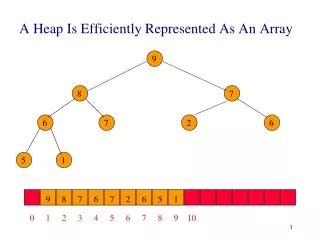
Matrix. The image is represented as a MATRIX of numbers . Matrix : A two dimensional array of numbers arranged in rows and columns . Each number represents the value of the image at that location. Voxel.

E N D
Matrix • The image is represented as a MATRIX of numbers. • Matrix: A two dimensional array ofnumbers arranged in rows and columns. • Each number represents the value of the image at that location.
Voxel • Each individual element or number in the image matrix represents a three dimensional volume element in the object, called a VOXEL.
Pixel • The VOXEL is represented in the image as a two-dimensional element called PIXEL - (picture element).
CT scan Generation موقعیت لامپ اشعه ایکس نسبت به آشکارساز تعیین کننده نسل سی تی اسکن است. نسل اول: سیستم های پرتو خطی (Pencil Beam) حرکت منبع اشعه ايكس و آشکارساز هر دو بصورت انتقال - دوران. نسل دوم: سیستم های پرتو بادبزنی باریک (Narrow Fan Beam) حرکت منبع اشعه ايكس و آشکارساز هر دو بصورت انتقال - دوران.
CT scan Generation • نسل سوم: سیستمهای پرتو بادبزنی پهن (Wide Fan Beam) حرکت منبع اشعه ايكس و آشکارساز هر دو بصورت دورانی. آغاز استفاده از فناوری حلقههای لغزنده (slip ring technology) • نسل چهارم: سیستمهای با حرکت دورانی منبع اشعه ايكس ، اما با آشکارساز ساکن.
CT scan Generation • نسل پنجم: سیستمهای مقطعنگاری رایانهای با پرتو الکترونی • نسل ششم: اضافه شدن حرکت مارپیچی یا اسپیرال (spiral) برای این نوع سیستمها، گام(pitch) قابل تعریف است. • نسل هفتم: استفاده از آرایههای آشکارساز چندردیفی (Multi Detector Array) معروف به MDCT.
CT scan Generation • امروزه پویشگرهای سیتی نسل هفتم بر اساس الگوی حرکتی سیستمهای نسل سوم کار میکنند، و سیستمهای نسل چهارم در واقع از رده خارج شدند. • لذا منشا پرتوها و آشکارسازها هر دو حرکت دورانی دارند. • همچنین با آمدن به بازار سیتیهای نسل ششم و هفتم با آرایهٔ ۶۴ برش، سیستمهای مقطعنگاری رایانهای با پرتو الکترونی تقریباً از صحنه حذف شدهاند، و امروزه بیشتر فقط برای پژوهش کاربرد دارند.
Dual Source CT scan • ارائه سي تي اسكن اسپيرال با دو منبع اشعه ايكس راهي است به سوي تشخيص تركيب شيميايي مواد در سي تي اسكن. • اين سيستم از دو منبه اشعه ايكس با انرژي 80 و 140 KVP بهره مي برد. • به طور معمول در سي تي اسكن 64 اسلايس بالاتر ديده مي شود.
كاربرد هاي فعلي : • سابتراكشن استخوان در تصاوير CTA • ارزيابي پرفيوژن ريوي • تشخيص نوع سنگ كليه • كاربردهاي در حال كار : • حذف يد از كبد براي توليد تصاوير مجازي بدون كنتراست • تشخيص ماهيت پلاك آترواسكلروتيك • ارزيابي پرفيوژن ميوكارد
The dual energycharacterization showsthat the calculus ofpatient 2 is a calcifiedstone, color-coded in blue(as cortical bone in theimage). In patient 1 thekidney stone can becharacterized as uric acid stone, color-coded in red.
[B] Corresponding angiographic image reconstructed from the same scan. There is embolic material in the lower and middle lobe arteries. [A] Color-coded sagittal MPR of the right lung showing normally perfused vessels in turquoise and embolized vessels in red.
[A] Axial image of both feet showing massive deformities and calcified masses [B] The red color-coding confirms uric acid in the masses, identifying them as gout tophi.