Download
1 / 15
160 likes | 395 Views
Meniscal tears, Osteoarthritis & Osgood- Schlatters Disease. By: Juliann Plimpton. Meniscal Tears. MOI/ Etiology Valgus force- adducts the knee- tears medial meniscus Varus Force- abducts the knee- tears lateral meniscus

E N D
Meniscal tears, Osteoarthritis & Osgood- Schlatters Disease By: Juliann Plimpton
Meniscal Tears MOI/ Etiology • Valgus force- adducts the knee- tears medial meniscus • Varus Force- abducts the knee- tears lateral meniscus • Weight- baring combined with rotary force while knee is flexed or extended • Londitudinal tear- knee is forcefully extended from a flexed position while the femur is internally rotated • Most tears happen as a combination of all these MOI: • Lateral rotation of the femur • Knee partially flexed • Foot firmly planted • Three zones: • Red-Red zone- outer 1/3 rich blood supply. Tears can heal over time when in this area • Red-White zone- middle 1/3 blood scares • White-White zone- inner 1/3 no blood circulation • Surgery is required when meniscal tears are in these zones.
Signs & Symptoms • Joint line pain and loss of motion • Intermittent locking. Giving way of the knee • Pain when athlete squats • Complaining of popping knee collapse • Swelling • Management • MRI • Arthroscopic surgery • Special Test • McMurray’s Meniscal test (p. 621) • Apley Compression test
Arthroscopic Surgery • The evaluation of a tear by inserting a blunt probe into the knee • Once evaluated the effected area is removed (meniscectomy) • Removed my cutting and sucking out tear
Techniques for Meniscal Tear Repair • Inside Out- curved guide tubes are used to direct a pair of long needles into the meniscus and out through a small incision in the back of the knee • Suture threads connected to the needles are then tied off out side of the knee • This works well but take note that a 11/2 to 2’’ incision needs to be made.
Cont. • Inside-In (T-Fix): includes meniscal staples and bioreabsorbable T- arrows. • sutures have an anchor, • Multiple sutures pairs are placed through long hollow needles • knot pusher instrument that securely snugs the meniscus down and provides an excellent repair.
Management • No weight baring for at least three weeks, with light-loading muscle exercises • After this physical therapy, maximal weight training not allowed until 2-3 months • Return to running and agility sports 3-4 months • With just the removal of a meniscal tear one recovers a lot quicker. • Back in play by 4-6 weeks.
Osgood-Schlatter & Larsen- Johansson Diseases • MOI of Osgood-Schlatters Disease- • Is an apophysitis characterized by pain at the attachment of the patellar tendon and tibial tubercle • Repeated avulsion of the patellar tendon at the apophysis • Common in adolescents • MOI of Larsen-Johansson Disease- • Occurs at the inferior pole of the patella • Due to excessive strain on the patella tendon • Signs and Symptoms • Swelling • Hemorrhages • Gradual degeneration of the apophysis • Athlete complains of severe pain when kneeling, jumping, and running • PT over anterior proximal tibial tubercle
CONT…. • Management- • Decrease stressful activity (6 months to a year) • Cylindrical cast • Ice • Isometric strengthening
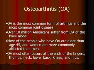
Osteoarthritis • MOI- chronic degeneration of the articular or hyaline cartilage • Wear and tear- to the pt. of exposing the bone • Repeated trauma to the joint, tendons, ligaments (running, cycling) • Signs and Symptoms • Pain brought by friction when in use • Stiffness • Tenderness • Creaking • Grating • Crepitus
Treatment • Hyalgan injections- purified Sodium hyaluronate • 5 injection over 5 weeks- works for 12 months • Glucosamine Sulfate- over the counter drug • Derivative of glycosaminoglycans found in articular cartilage
Looking beyond the Drugs… • Worse case… Surgery. • Three methods: • Arthroscopy • Osteotomy • Arthroplasty http://www.edheads.org/activities/knee/
References • Avery, Lincoln. (2006). “The Meniscus: Shock Absorber for the Knee”. http://www.orthoassociates.com/meniscus.htm. • Grainger, Rebecca; Cicuttini, Flavia. (2004). “Medical management of osteoarthritis of the knee and hip joint”. The Medical Journal of Australia. 232. http://www.mja.com.au/public/issues/180_05_010304/gra10763_fm.html. • Prentice, E. William: “Musculoskeletal Conditions. Arnheim’s Principles of Athletic Training 12th ed.: 585, 2005. • “Surgical Treatment of Osteoarthritis of the knee”. (2003). http://orthoinfo.aaos.org/fact/thr_report.cfm?Thread. • http://www.coachroblowe.com/injuries-lower-osgood-schlatters.jpg. • http://www.radiology.vcu.edu/Peds%20COTW/2006/03-30-06/lat.jpg.>