Download
1 / 53
590 likes | 996 Views
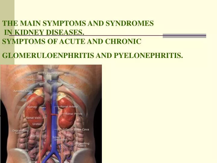
THE MAIN SYMPTOMS AND SYNDROMES IN KIDNEY DISEASES. SYMPTOMS OF ACUTE AND CHRONIC GLOMERULOENPHRITIS AND PYELONEPHRITIS.

E N D
THE MAIN SYMPTOMS AND SYNDROMES IN KIDNEY DISEASES. SYMPTOMS OF ACUTE AND CHRONIC GLOMERULOENPHRITIS AND PYELONEPHRITIS.
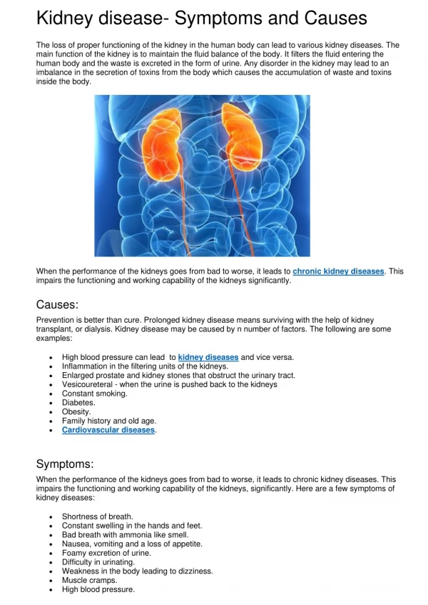
What do the kidneys do?1.remove toxic waste products 2. remove excess water and salts 3. take part in controlling your blood pressure 4. produce erythropoietin (epo for short) which stimulates red cell production from the bone marrow - you get anaemic without this 5. help to keep calcium and phosphate in balance for healthy bones 6. maintain the blood in a neutral (non-acid) state
main pain in lumbar region, urination disoders, change of urine, oedema. secondary chills, headache, dizziness, vision deranged, heart pain, skin itching, loss of appetite, nausea, vomiting , fever. Signs, which point on kidney affection
Pain in lumbar region Pain is caused by: stretching of kidney capsule, spasm of urethers, inflammation of peryrenal cellular tissue, kidneys infarction. Intensity of the pain feelings can be different – from dull, boring pain (at acute and exacerbation of chronic pyelonephritis) to sharp, very severe pain with an irradiation along ureters, in a groin, in genitals, in the front surface of thigh (nephrocolic which arises up as a result of ureter corking with a stone, at his bend, at the trauma of kidneys, kidneys infarction).
Urination disoders Oliguriya(decreasing of urine excretion of less than 500 ml per day). Anurya(complete stop of urine excretion - symptom of acute kidney insufficiency, mechanical obstacles of urine passage (stone, tumour). Nycturia(advantage of nightly diuresis above daily ( normaly 1:2); Dysuriya – (painful urination) Polakiuriya – (frequent urination, which combines with polyuria at chronic nephritis, cystitis). Isosthenuria(At disorders of dilution and concentration function of kidneys specific gravity is 1010-1011)
Oedema are one among the main symptoms of renal pathology. Often oedema is the first sign of such diseases. Renal oedema can suddenly develop as well as disappear. As a rule they are located on the face especially on eyeleads (where the subcutaneous fat tissue is more loose ). Sometimes oedema is equally distributed all over the body (anasarka). Oedema have mild consistency, deep elevation of skin is present even in slight pressing on it. Oedema can spread on internal organs and cavities (profound oedema) with accumulation of transsudate in serous cavities — pleural, abdominal and pericardial cavities.
Uric syndrome. • moderate proteinuria (from 100 mg up to 3,5 g a day), • red blood cells in the urine more than 1106/L per day (erythrocyturia), • leukocyturia - more than 1106/l per day), • custs in urine, • bacteriuria, • dischargingof salts and their cristals with urine • cells of renal and transitional — epithelium and other elements of pathological both organized and non- organized renal sediments, • The leading symptom in uric syndrome is proteinuria (albuminuria).
Nephrotic syndrome. • A massive leak of protein (albumin) into the urine (proteinuria) (more than 3,5 gr per day). • A low blood level of albumin due to the large amounts lost in the urine (hypoproteinemia mostly because of hypoalbuminemia). • An increased level of cholesterol in the blood (hyperlipidemia). • Retention of fluid in the body (edema) causing swelling. • Hypercoagulation.
Renal arterial hypertension. This is symptomatic hypertension caused by affection of kidneys or renal vessels with following disorders of blood pressure regulation. • Elevation of blood pressure is caused by 3 mechanisms: 1.sodium and water retention, 2.activation of pressor system, 3.inhibition of depressory mechanisms.
Acute nephritic syndrome. It is characterized by the abrupt onset (days) of haematuria with red blood cells, casts or dysmorphic red blood cells appearing in the urine, proteinuria, renal impairement (oliguria, uremia, raised urea and creatitine), hypertension due to water and salt retention, edema (usually periodical).
What is glomerulonephritis? • Diffuse glomerulonephritisis the general infectious-allergic disease with prevalent and primary envolvement of nephrone glomerular apparatus into the pathological process. • Glomerulonephritis is a type of glomerular kidney disease in which the kidneys' filters become inflamed and scarred, and slowly lose their ability to remove wastes and excess fluid from the blood to make urine. • Types of glomerulonephritis include kidney • disease of diabetes, IgA nephropathy, and • lupus nephritis.
Classification (by L. Pyrih) • The following forms of glomerulonephrites are determined • I. Acute diffuse glomerulonephritis : • uric syndrome; • nephrotic syndrome (mostly haematuria, hypertension and edema are present). • II. Subacute fulminant glomerulonephritis. • III. Quickly progressing glomerulonephritis. • IV. Chronic glomerulonephriis: • 1st type: primary chronic, secondary chronic. • 2. syndromes:uric, nephrotic. • 3. stages: • non-hypertensive, hypertensive; • chronic renal failure; • 4. phase:exacerbation, remission.
Acute glomerulonephritis(AG, lat. - glomerulonephritis аcutа). • This is acute immune inflammatory lesion of kidneys parenchima with primary damage of glomerules and following involvement of all renal structures into the pathological process.
Ethiology and pathogenesis AG develops 2-3 weeks after acure infectious disease (tonsillitis pharyngitisskarlet fever more rarely —etysipelasetc.) caused by -haemolyticstreptococcus, group A or by otherbacteriological agents (pneumococus, staphylococcus, viruses). The process develops as a hyperergic reaction of sensebilized organism. Administration of serum preparations, other medical preparations, vaccination, penetration of toxic substances into the organism, deranged venous outflow from kidneys may be the causes of AG. Provoking factors are overooling, dump weather, traumas.
1. Enlarged and hyperaemic glomeruli; 2.Ischaemia of the glomeruli; 3. Fibrinoid swelling of the capillary walls, proliferation of their endothelium; 4. Accumulation of coagulated proteinous exudates in the space between the capillary loops and the glomerular capsule, blood stasis, thrombosis of the capillary loops, and hemorrhages. Pathological anatomy
This symptoms are combined in followingsyndromes: hypertension, uric syndrom, edema • Uric syndromeispresence of protein in the urine, blood formed elements, casts. • Even at the beginning of the disease urine colour is changed because of blood admixtures. Urine has the colour of “meet wastes”. Erythrocytes quantity at the peak of the disease is about 100—200 in a vision field. • Hypertension occurs due to activation of renin-angiotensin-aldosterone and sympathoadrenal systems as well as water and salt retenstion in the organism and inhibition of depressor systems. • Blood pressure elevates from insignificant level till 180-200 mm Hg (systolic) and 120 mm Hg (diastolic one). • Main complaints of these patients are: headache, heaviness in the head, dyspnea, palpitation, nausea and sometimes — periodical imparement of vision.
Data of general inspection: • patient’s appearance is quite specific and it is called “facies nephritic a”: • skin paleness, • swelling of the face and eyeleads edema under the eyes, • Patient’s condition is heavy, his posture in bed may be forced — sitting or semirecumbent.
:Palpation: pulse is full, dull and slow. Apex beat is intensified and displaced leftward.Percussion: may reveal fluid in pleural cavity, lung congestion and displacement of left heart border leftward. Auscultation: bradicardia, I heart sound is weakened, systolic murmur over heart apex, accentuation of the II heart sound over the aorta.In severe cases especially in left ventricular hyperthrophy hallop rhythm occurs. Vesicular breathing is heard over the lungs.
Laboratory methods of examination: • Total blood count: moderate leukocytosis anaemia accelerated ESR. • Вiochemical blood analysis — increased content of seromucoid, sialic acids fibrinogen, C-reactive protein, antistreptoliase, immune complexes, 2- і -globulines. In heavy cases oliguria, decreased urine density, nicturia. In resilience of edema (period of recovering) amount of urine increases and ability of kindeys for concentration raises.
Instrumental methods of examination: • Eye grоunds:narrowing of arterioles, dilatation of veins, sometimes - aedema of ophthalmic nerve, hemorrhage into the retina. • ECG— overloading and hyperthrophy of the left ventricle decreased voltage of Р і R, depression of ST interval, T is low of biphasic. • X-ray examinationof the chest: probable presence of fluid in pleural cavity signs of lung congestion, enlargement of the right ventricle.
Treatment: • bed regimen for 1-15 months dry and warm ambient temterature. • Diet № 7, limitation of salt ( to 05-15 gr a day) liquid. • Medicamentous treatment — antibiotics for 7-10 days (penicillin or semisynthetic penicillins). • Hypotensive agents: sedative (valerian mothewort bromine tranquilizers) antiadrenergic preparations (apressinclonidine metildopum, -bloquers) in combination with saluretics, losartan (angiotensin converting enzyme inhibitor), perypheric vasodilatators. • Diuretics (in edema, heart failure, hypertension). • Pathogenetic therapy — immunodepressants glucocorticoids agents that improve haemosthasis and microcirculation (indometacin tiklopidine, curantyl heparin).
Chronic glomerulonephritis • (glomerulonephritis chronica, ChG) is inflammatory process in renal glomeruli degeneration of canalicular epithelium and progressive development of connective tissue that resulting in secondary shrinked kidney. • Ethiology and pathogenesis. Often ChG develops after acute glomerulonephritis. If information about AG in patient’s anamnesis is abcent than thay speak about primary form of ChG
Clinical picture. • In unhypertensive stage: general weakness, quick fatigue, dull boring pain in lumbar region, change of urine colour, edema below eyes and on the face, on the legs. • In hypertensive stage: the same plus headache, dizziness, periodical nassal bleeding, dyspnoe, nictural dyspnoe, palpitation.
Data of inspection: patient’s condition is satisfactory in remission but in exacerbation may be severe. Skin is pale, edema are visible on the face (below the eyes (facies nephritica). Sedimentation of uric acid cristals is possible on the skin. Inspection of precordial region: displacement of apex beat leftward from the left midclavicular line. In dilatation of the left ventricle apex beat becomes diffuse.
Palpation:Apex beat is intensified, diffuse and displaced leftward. Percussion: the left border of relative heart dullness is displaced leftward from the left midclavicular line in V interspace. Palpation of organs of abdominal cavity: is painful in epigastrium and above the large bowel. In the case of right ventricular failure the liver is enlarged.
Auscultation: in patients with left ventricular failure moist rales may be heard in lover parts of the lungs (because of lung congestion). Heart soungs are intensified but later become weakened, accenttuation of the II sound is heard over the aorta, systolic murmur over the apex. In terminal stage pericardial friction sound may be haerd on the hart basis, along the left sternal border and in zone of absolute heart dullness. Blood pressure is elevated up to 200/120 mm Hg. If heart failure develops systolic pressure may decrease but diastolic one is steel high.
Chronic glomerulonephritis with nephrotic syndrome is manifested by decreased diuresis, edema and changes in the blood and urine: hypo- and dysproteinaemia (hypergammaglobulinaemia, hypoalbuminaemia), hyperlipidaemia, proteinuria more than 3,5 gr/l, casts in urine (hyaline, granular and rarely - ceraceous (waxy). Respiratory infections are frequent because of immunodepression that provoke exacerbation of ChG.
Eye grounds: retinal arteriosclerosis hemorrhagias focci of degeneration and affections of n. ophthalmicus (neuroretinopathy). • In the stage of renal failure patient’s condition is heavy. Forced posture (sitting). Deranged conscioussness, sometimes coma. Main complaints are nausea, vomiting, skin dryness and itching, deranged vision, oliguria or anuria. • Laboratory examination. Changes in urine: compensatory poliuria, nikturia. Zimnitsky’s test show hypoisosthenuria, nicturia. Urine density decreases and become monotonous hypoisosthenuria (1009-1012). Creatinine and urea content in the blood may be normal. • Proteinuria from insignificant till 3-10 gr/l. Casts — hyaline granular and ceraceous.
Laboratory examination. • Changes in urine are folowing: compensatory poliuria, nikturia. Simnitsky’s test show hypoisosthenuria, nicturia. Urine density decreases and become monotonous hypoisosthenuria (1009-1012). Due to poliuria products of nitrogen metabolism is possible to evacuate from the organism that is why kreatinine and urea content in the blood may be normal. • Proteinuria develops. Its degree may be from insignificant till 3-10 gr/l. It depends on patient’s diet, physical loading, overcooling etc. On urine sediments there are casts — hyaline granular and ceraceous.
Treatment is perormed taking into account clinical variant of the disease, its phase and stage. Patient should avoid overcoolings, physical loadings, psychoemotional stresses. In exacerbation long-standing treatment is indicated. Diet № 7, limitation of salt intake to 10 gr a day in nephrotic variant - 6-8 gr a day.
Treatment • In exacerbation long-standing treatment is indicated. • Diet № 7, limitation of salt intake to 10 gr a day in nephrotic variant - 6-8 gr a day. • Medicamentous treatment: In unhypertensive stage - corticosteroids, cytostatics, aminoquinoline derivatives. In hypertension hypotensive drugs also should be prescrobed (-bloquers methyldopum, apressin etc.). • Diuretics (furosemide ethacrynic acid, hydrochlorothiazide) in edema; • Anticoagulants and antiaggregants (heparin curantyl indometacin) • Hemosorption, plasmapheresis. • If infection develops than antibiotics are administered. • Nephrotic syndrome is indication for administration of prednisolone 30-80 mg a day, azathioprine – 100-150 mg a day, heparin – 10000-15000 Un, aspirin – 0,25 gr. • In remission stage – sanatorium-resorting treatment is useful with warm and dry climate, mineral waters. • If high risk of renal failure and exaggerated asotaemia are present it is necessary to perform chronic hemodialisis and transplantation of kidney.
Prognosis.Duration of patient’s life depends on clinical form of ChG and functional condition of kidneys. In the stage of chronic renal failure patients are disable.Prophylaxisincludes in-time treatment of acute and chronic focci of infection as well as treatment of exacerbations, dyspancery observation.
Pyelonephritis • Pyelonephritis is a bacterial infection of one or both kidneys. • Ethiology:E. Coli, streptococci, staphylococci, proteus and differrent bacterial assotiations, sometimes it may be caused by viruses • Provoking factors: disorders of urine outflow (congenital anomalias, stones, obstruction, pareses, paralises etc.); metabolic disorders (diabetes mellitus, gout); iatrogenic diseases (catheterization, cystoscopy); immunodepression (chronic diseases, focci of infection, overcooling).
Pathogenesis: Penetration of microorganisms to calicules and renal interstitium by hematogenic or ascending ways (thrugh urinary tract). • Classification:unilateral and bilateral, acute and chronic pyelonephritis, primary and secondary. • Primary pyelonephritis occurs when morphological changes in urinary tract are abcent. • Secondary pyelonephritis occurs when anomalias of kidneys and urinary tract are present which cause disorders of urine outflow (narrowing of ureters or urethra, stones nephroptosis adenoma of prostatic gland etc.). • Acute pyelonephritis devides on serous and purulent.
Clinical pattern. • Acute pyelonephritis (AP) starts from elevation of body temperature up to 38-39С chills headache unilateral or bilateral dull pain in lumbar region dysuria. Nusea, vomiting, myalgias and arthralgias are possible. Patient’s condition is heavy, toxic shock may develop. Respiration is frequent, vesicular. Tachicardia is present. • Tongue is dry and coated. Herpes labialis. Urination is frequent painful sometimes urinary retention develops. Pasternatsky’s symptom is positive on the affected side.
Urine relative density is decreased (1012-1015) Poliuria is possible. • Urine reaction is acid. Leukocyturia and bacteriuria are typical. Non-significant haematuria and proteinuria (till 1 gr/l), casts in urine (hyaline granular and epythelial). • Nechiporenko’s and Addis-Kakovsky’s tests are positive.Sometoimes urine is of alkaline reaction and of unpleasant smell, becomes cloudy, salt sediments and purulent flakes are present is it. • Microscopic examination: leukocytes, granular custs and erythrocytes on all fision fields.
Treatment: • Вed mode; diet enriched with milk, vegetables and fruits. Alkohol, spicy food preserved food coffee are prohibited. Liquid 3 liters a day (wild rose decoction, mineral water). • Antibioticotherapy: а) penicillin, semisynthetic penicillicnes, hentamycin, claforan ets., b) nitrofurantoin derivatives: nitrofurantoin (0,1 g 3—4 tablets a day 7-10 days), biseptol-480 (2 tablets 2-3 times a day 2-3 weaks). • Spasmolithics and diuretics. • In heavy cases catheterization, lawage of calices and bladder with desinfectant solutions.
Chronic pyelonephritis • Chronic pyelonephritis (pyelonephritis chronica, ChP ) is a chronic non-specific inflammation of renal interstitium and calicular mucosa with following affection of renal vessels. • Ethiology and pathogenesis. Chronic pyelonephritis may be the result of not effective treatment of acute pyelonephritis. Provoking factors of ChP: urine congestion, inflamatory diseases of female sexual organs, appendicitis, urolithiasis, diabetes mellitus, chronic intoxications, inhibition of immune system. • The main way of penetration of infection is ascending in disorders of urine outflow (kidney stones, anomalias and strictutes (narrowings) of urinary tract tumors pregnancy etc.). Infection spreads from renal calices to the renal parenchyma.
Clinical pattern. There are latent, recidiving, hypertensive, anaemic and asotaemic forms of the disease. Main complaints are elevation of body temperature, chills, pain in the projection of one or two kidneys, headache general weakness fatique dysuria. If the patient develop renal failure, his appetite is lost, nausea, vomiting, thirst and metheorismus are present.
Blood analysis: anaemia leukocytosis neutropenia and lymphopenia thrombocytopenia accelerated ESR. • Urine: its density decreases to 1003-1005 (hypoisosthenuria). Mild proteinuria (to 1 gr/l), leukocyturia. • Nechiporenko’s and Kakovsky-Addis’ tests data: leukocyturia prevails before erythrocyturia. • Bacteriologic examination of the urine reveales marked amount of bacteria. • Zimnitsky’s test at the beginning of the disease reveales hyposthenuria and later hypoisosthenuria. • Kreatinine and urea clearance are decreased. The level of blood kreatinine and urea rise. • X-ray, ultrasonic examination and computer tomography show distension and deformation of renal calices, asymethry, shrinked kidneys.
Treatment: to avoid overcoolings and viral respiratory infections; diet № 7 (in exacerbation). In anaemia - food rich on iron (strawberries apples); grapes, melon and water-melon. • Antibiotics in 10-12- days courses with breaks for 2-3 weaks. nalidixic acid derivatives — negram nevigramone; nitrofurantoins biseptol sulfa-drugs of short action (ethazol urosulfane). • Vitamins of B group, ascorbic acid antihistamine preparations and spasmolitics. • In the case of hypertesion salt intake should be limited to 4-6 gr a dayhypotensive drugs should be prescribed. • In remission sanatory-resorting treatment is useful . • Prognosisis favorable in active treatment and abcense of complications. • Prophylaxis of ChP means treatment of chronic infection focci as well as inflammation of urogenital tract, observation of rules of aseptics and antiseptics during instrumental examinations (cystoscopy, catheterization etc.).
Simple renal cysts demonstrated at MRI The patient had one cyst in each kidney.a) T1-weighted image.b) T2-weighted image. MRI clearly delineates renal cysts. On T1-weighted images the content is signal poor often with a clearly delineated wall, whereas on T2weighted images it is signal intensive, sometimes with edge enhancement
Figure 32.Adult autosomal dominant polycystic kidney disease demonstrated at intravenousurography. Both kidneys are enlarged with irregular contours. The pyelocalyceal systems are splayed and deformed.