Download
1 / 39
390 likes | 598 Views
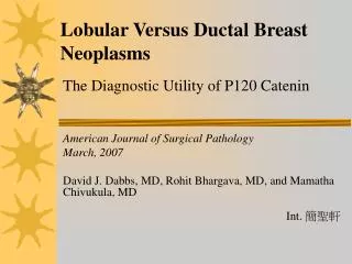
Lobular Versus Ductal Breast Neoplasms. The Diagnostic Utility of P120 Catenin. American Journal of Surgical Pathology March, 2007 David J. Dabbs, MD, Rohit Bhargava, MD, and Mamatha Chivukula, MD. Int. 簡聖軒. Introduction.

E N D
Lobular Versus Ductal Breast Neoplasms The Diagnostic Utility of P120 Catenin American Journal of Surgical Pathology March, 2007 David J. Dabbs, MD, Rohit Bhargava, MD, and Mamatha Chivukula, MD Int. 簡聖軒
Introduction • The diagnostic discrimination of ductal versus lobular neoplasms is important in several circumstances for therapeutic reasons. • Diagnostic reproducibility of lobular versus ductal lesions, based on histology alone, is less than optimal. • It is important to correctly diagnose the specific types of breast neoplasia so that appropriate risk assessment and prognostic and predictive tests will be performed and appropriate therapies instituted.
Introduction • The broad categories of ductal and lobular neoplasias of the breast represent the majority of important breast lesions, but there are striking differences in morphology, molecular alterations, and treatment strategies for each category. • The proper distinction between atypical lobular hyperplasia, lobular carcinoma in situ and low-grade ductal carcinoma in situ is critical for patient management.
Introduction • Low-grade ductal carcinoma in situ Surgical excision with proper margins and perhaps radiation therapy and continuous follow-up. • Lobular carcinomas I : Lumpectomy + ALND + C/T II, III : Mastectomy + ALND + C/T IV : C/T + selective H/T
Introduction • The pathologic diagnosis of metastatic breast carcinoma remains a challenge even today. • A plethora of antibodies in the diagnostic armamentarium, and less than a handful of antibodies are useful in identifying breast carcinomas in metastatic sites. • Gross cystic disease fluid protein fraction 15 (GCDFP-15) • Hormone receptors estrogen and progesterone (ER/PR) • Cytokeratin 7 (CK7) • Mammaglobin antibody
Introduction • In the breast, E-cadherin is useful to distinguish between ductal and lobular neoplasia. • Ductal : Cell membrane, E-cadherin (+) • Lobular : E-cadherin • E-cadherin is present in most epithelia, and therefore is not specific for diagnostic use in the setting of metastatic carcinomas.
Introduction The E-cadherin complex is composed of the transmembrane E-cadherin protein and α,β,γ, and P120 catenins (ctn) which anchor the E-cadherin protein to the cytoplasmic actin filaments.
Introduction • We and others have recently discovered a characteristic cytoplasmic redistribution of P120ctn in lobular neoplasia. • Ductal : dominant membrane immunostaining pattern of P120ctn • Lobular : diffuse cytoplasmic P120ctn immunostaining pattern
Introduction • In this study, we survey neoplastic ductal and lobular lesions of the breast with P120ctn to determine its utility for the surgical pathologist. • In addition, we survey a broad variety of other carcinomas to determine whether the P120ctn cytoplasmic immunostaining pattern seen in lobular neoplasia may be useful in the diagnosis of metastatic lobular carcinoma.
Method • Formalin-fixed ; Paraffin-embedded • Invasive lobular carcinoma : 64 cases Invasive ductal carcinoma : 62 cases • These cases were retrieved from the archives of the Department of Pathology, Magee-Women’s Hospital. • For the study of neoplastic breast lesions, each case was examined by a hematoxylin/eosin section, E-cadherin, and P120ctn independently.
Method • Each tumor was classified as lobular or ductal on the basis of standard histologic criteria. • Cell membrane immunostaining for E-cadherin was considered to be a ductal immunophenotype. Complete lack of the membrane immunostaining was considered to be a lobular immunophenotype. • Each case was independently reviewed for the pattern of cellular immunostaining for P120ctn.
Method • Ductal immunophenotype : Cell membrane immunostaining for P120ctn of any intensity, without cytoplasmic immunostaining. • Lobular immunophenotype : Diffuse cytoplasmic immunostaining without any appreciable cell membrane accentuation for P120ctn.
Method • To survey a broad array of carcinomas for the presence of cytoplasmic P120ctn immunostaining, tissue microarrays (TMA) were used. • TMAs were fixed in formalin, paraffin embedded and subject to the same immunostaining procedure as the breast tissue sections described above. • A summary of the TMA components with detailed immunostaining scores is listed in Table 1.
Method • P120ctn Stain • C : Cytoplasmic • C+M : Cytoplasmic and Membrane • M : Membrane • Tissue microarrays (TMA) • Weak: 1+ staining in any percentage of cells or 2+ staining in <10% of cells • Moderate: 2+staining in >10% of cells or 3+ staining in <10% of cells • Strong: 3+ staining in >10% of cells
Result • Lobular neoplasia There was a lack of E-cadherin membrane staining and there was a diffuse, intense cytoplasmic P120ctn immunostaining. • Ductal carcinomas Distinct membrane accentuation pattern of P120ctn was noted.
Invasive lobular carcinoma, hematoxylin/eosin B. P120ctn: invasive lobular carcinoma with diffuse cytoplasmic stain
C. Comparison of membranous P120ctn staining in normal duct to the cytoplasmiconly staining of lobular carcinoma D. Negative E-cadherin of the same case in (C), membranous pattern in normal duct
Result • P120ctn was a diagnostic aid in challenging case. • Sometimes, the immunostaining of myoepithelial cells with E-cadherin in lobular neoplasia may be a challenge to interpret.
Duct epithelial hyperplasia Or Atypical lobular hyperplasia
P120ctn shows intense cytoplasmic immunostaining of ALH cells appearing in the lobule. Membranous immunostaining
E-cadherin result of this same case is less conspicuous, but shows lack of membrane immunostaining of ALH cells. Myoepithelial cells are stained with E-cadherin. Shows immunostaining of myoepithelial cells with smooth muscle myosin heavy chain in the section adjacent to (E).
Low-grade ductal neoplasia or Lobular neoplasia with microcalcification?
P120ctn confirms lobular neoplasia with characteristic intense, diffuse cytoplasmic immunostaining.
Result • The E-cadherin and P120ctn patterns defined the ductal or lobular types. • Classic and pleomorphic variants of lobular carcinoma showed identical patterns for P120ctn
Pleomorphic variant of lobular carcinoma, hematoxylin/eosin . Diffuse cytoplasmic pattern for P120ctn.
Result • In the TMA survey, some rectal (9/63) and gastric (3/72) adenocarcinomas • Dominant cytoplasmic P120ctn immunostaining pattern • Similar to lobular neoplasms. • These rectal and gastric carcinomas were all of the diffusely infiltrating pattern with various degrees of mucinous signet ring cells, mimicking the appearance of lobular carcinoma • None of the other carcinomas demonstrated the exclusive diffuse cytoplasmic P120ctn immunostaining pattern.
Gastric carcinoma Rectal carcinoma • The rectal (A) and gastric (B) carcinomas of the diffusely infiltrating pattern mimic the appearance of lobular carcinoma • All had strong cytoplasmic P120ctn.
Discussion • Sarrio et al demonstrated that P120ctn had a cytoplasmic localization in ALH, LCIS, and invasive lobular carcinoma. • We also documented P120ctn cytoplasmic immunostaining in ALH, and found it to be somewhat easier to interpret than negative E-cadherin in early cases of ALH where duct epithelial hyperplasia was in the differential diagnosis.
Discussion • Nearly 90% of diffusely infiltrating gastriccarcinomas showed cytoplasmic P120ctn whereashalf of these cases also lost membrane P120ctnimmunostaining. • A similar distribution of E-cadherinand P120ctn was seen in colorectal carcinomas. • Skoudy et al using fresh frozen tissue and a differentP120ctn antibody, found less than half of colon adenocarcinoma cells had cytoplasmicpositivity.
Discussion • In our study, the diffusely infiltrating rectal and gastric tumors which morphologically mimic invasive lobular carcinoma, also demonstrated diffuse cytoplasmic immunostaining for P120ctn in every cell. • P120ctn alone would not be useful to diagnostically separate lobular carcinoma from these diffuse-type rectal and gastric carcinomas. • P120ctn may be a useful part of a diagnostic panel of antibodies in the workup of a case in which breast carcinoma is in the differential diagnosis. • An immunopanel that includes gastrointestinal markers and estrogen receptor would be necessary to definitively make this distinction.
Discussion • A diffuse strong cytoplasmic P120 immunostaining pattern in a dyscohesive carcinoma should raise the possibility of lobular breast carcinoma. • Additional antibodies that would be useful to distinguish lobular carcinoma from the diffusely infiltrating variants of rectal and gastric carcinoma would include estrogen receptor, GCDFP-15, mammaglobin, cytokeratins 7 and 20, and CDX-2 transcription factor.