Download
1 / 1
10 likes | 98 Views
This study explores the vascular regeneration in rat liver after partial hepatectomy, focusing on the growth patterns and fenestration of sinusoids. Scanning electron microscopy reveals insights into how the vasculature compensates for the removed liver section. The control group and experimental group were compared to understand the significance of weight changes and vascular regrowth differences. Further research aims to delve deeper into this complex circulation and analyze the growth factors associated with the regeneration process.

E N D
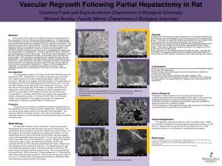
Vascular Regrowth Following Partial Hepatectomy in Rat Courtney Frank and Kayla Anderson (Department of Biological Sciences) Michael Bentley, Faculty Mentor (Department of Biological Sciences) • Results • A statistical t-test was performed to determine the significance between the starting weight and ending weight for the control group and the experimental group. There were no significant differences between the body weights of the control group and the experimental group after the two day period. • A statistical t-test was performed to determine the significance between the control group mean weight and the partially hepatectomized group mean weight. There was no significant difference between the weights of the two groups after the two day period. • Upon dissection of the hepatectomized rats after two days, re-growth was not visible in the resected median lobe, but instead in the remaining lobes. • According the SEM images, fenestrations were visible amongst the vessels. Abstract The purpose of this study is to examine the growth and regeneration of the vasculature of the liver following a partial hepatectomy. The regeneration of the hepatocytes of the liver has been studied extensively, but little attention has been directed towards the regeneration of the supportive vasculature. The vasculature of the liver is highly complex. The liver receives blood from hepatic arteries as well as the hepatic portal vein; therefore, the blood supply in the capillaries (hepatic sinusoids) is a mixture of arterial and portal blood. It is currently unknown how this complex circulation is reestablished in relationship to the regeneration of hepatocytes. In order to perform this study, a group of rats were assigned to one of two groups. The first group was composed of rats that were given the experimental surgery and the other group was given the sham surgery. The vasculature was prepared for viewing two days after the hepatectomy. The vasculature was then viewed under the scanning electron microscope. After viewing the vasculature, it appears that there is fenestration of the sinusoids in the experimental group and in the control group, with the control group having greater fenestration. However, further investigation is underway. In conclusion, it seems the vasculature in the livers of the experimental group are compensating for the portion of the liver that was removed. • Conclusions • After two days, the experimental group livers show few fenestrations amidst the vessels and hepatocytes. • After two days, the control group livers show fenestrations amidst the vessels and hepatocytes. • At this time, the scanning electron micrograph images of the hepatectomized liver show vasculature varying in diameter and consistency. The vasculature of the experimental liver appears more attenuated than the normal liver. Introduction The regenerative capacity of the liver is well known (Michalopoulos and DeFrances, 1997). Madrahivmov et al (2006) showed survival of rats with 90% surgical resection by using an anatomically based, vessel-oriented procedure. Although much research has been focused on the regeneration of the hepatocytes of the liver (Michalopoulos and DeFrances, 1997), not much has been focused on the regeneration of the vasculature of the liver. It has been shown that angiogenesis, the formation of vessels, occurs with liver regeneration (Drixler et al, 2002). However, the structural aspects of the growth and regeneration of the vessels of the liver has not been shown. Consequently, it is uncertain whether the blood supply to the regenerating tissue is mainly arterial, portal, or a mixture of both. Also, it is not known whether the arrangement of the hepatic sinusoid with the regenerated hepatocytes is ordered in a similar fashion to normal tissue. Pictures 1-4:These pictures show the fenestration in the sinusoids of the rat liver. Starting in the upper left corner, each picture is of the same sinusoid at varying magnifications. • Future Research • Research on the re-growth of the vasculature will be continued. • Additional scanning electron micrographs will be taken of the control and hepatectomized livers for examining the re-growth of the vasculature. • Amounts of different vasculature growth factors may also be studied to determine where the re-growth is located. • The fenestrations in the regenerated livers may be measured to analyze the experimental versus control livers more efficiently. Purpose The purpose of this study is to examine the growth and regeneration of the vasculature of the liver following a partial hepatectomy. Scanning electron microscopy will be used to examine the regrowth of the vasculature. The scanning electron microscope will allow us to view the vasculature and determine whether or not the vasculature in the regeneration tissue is similar to normal liver size. Acknowledgements This research project was funded by a URC Foundation Grant. Thanks to Dr. Knoblich for teaching us her surgical techniques and allowing us to use her lab. Thanks to Brent for all the help in the animal care room. Most importantly, thank you to Dr. Bentley for his dedication and support. Methodology Ten male WKY rats were used in this project. Five were used as an experimental group and the other five were used as the control group. The experimental group underwent a partial liver hepatectomy (lobe removal). For the surgeries, the rats were anesthetized by isofluorane inhalation. For the experimental rats, an incision was made and the middle lobe was clamped, sutured with absorbable thread, and removed. For the control rats, the same incision was made, but no liver manipulation occurred. Rimadyl was injected under the skin after surgery. Two days post procedure, the liver was extracted from the rat. The rats were anesthetized with inactin. Heparin was also injected into the rat before the extraction. The hepatic portal vein was cannulated with polyethene tubing. Through the tubing, an infusion of .9% saline solution was administered to drain the blood. The rat was euthanized by pneumothorax. After euthanasia, 2.5% glutaraldehyde in 0.1 M phosphate buffer was infused through the cannula. The liver was removed, post fixed for one hour in 1% osmium tetroxide, dehydrated in acetone, critical point dried, and analyzed by scanning election microscopy. • References • Drixler TA, Vogten MJ, Ritchie ED, van Vroonhoven TMJV, Gebbink MFBG, Voest EE, Rinkes IHMB (2002) Liver regeneration is an angiogenesis-associated phenomenon. Ann Surg 236:703-712. • Madrahimov N, Dirsch O, Broelsch C, Dahmen U (2006) Marginal hepatectomy in the rat: from anatomy to surgery. Ann Surg 244:89-98 • Michalopoulos GK and DeFrances MC (1997) Liver regeneration. Science 276: 60-66. Pictures 5-8: Pictures of control sinusoids showing fenestration.