Download
1 / 34
340 likes | 467 Views
The Cardiovascular System Chapter 11a. The Cardiovascular System. A closed system of the heart and blood vessels The heart pumps blood Blood vessels allow blood to circulate to all parts of the body

E N D
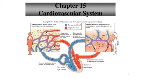
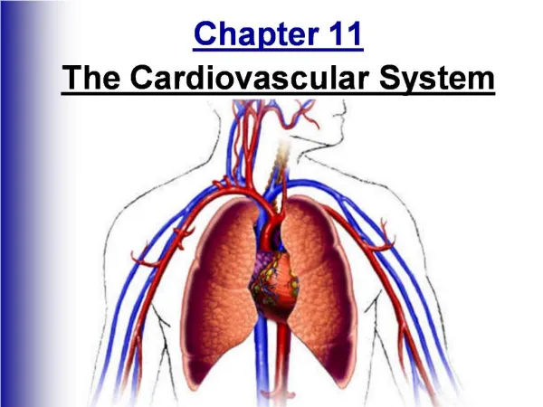
The Cardiovascular System • A closed system of the heart and blood vessels • The heart pumps blood • Blood vessels allow blood to circulate to all parts of the body • The function of the cardiovascular system is to deliver oxygen and nutrients and to remove carbon dioxide and other waste products
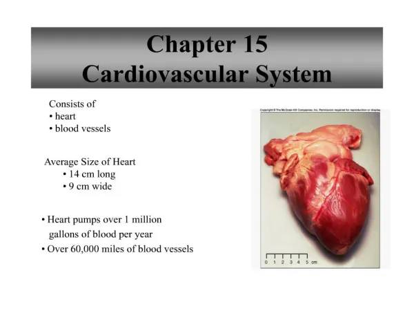
The Heart • Location • Thorax between the lungs • mediastinum • Pointed apex directed toward left hip • At 5th rib • Posteriosuperior = base (top) • At 2nd rib • About the size of your fist
The Heart Figure 11.1
The Heart: Coverings • Pericardium – a double serous membrane • Visceral pericardium • Next to heart • Parietal pericardium • Outside layer • Anchors heart to diaphragm & sternum • Serous fluid fills the space between the layers of pericardium • Decreases friction
The Heart: Heart Wall • Three layers • Epicardium • Outside layer • This layer is the parietal pericardium • Connective tissue layer • Myocardium • Middle layer • Mostly cardiac muscle - twisted • Endocardium • Inner layer • Endothelium – continues into vessels
External Heart Anatomy Figure 11.2a
The Heart: Chambers • Right and left side act as separate pumps • Four chambers • Atria • Receiving chambers • Right atrium • Left atrium • Ventricles = pumps • Discharging chambers • Right ventricle • Left ventricle Figure 11.2c
Septum – divide longitudinally • interatrial • Interventricular • Double pump • Right – pulmonary • Oxygen-poor blood through superior & inferior vena cava & out pulmonary trunk (right & left arteries) lungs • Left – systemic • Left ventricle is more muscular
Blood Circulation Figure 11.3
The Heart: Valves • Allow blood to flow in only one direction • Four valves • Atrioventricular valves – between atria and ventricles • Bicuspid valve (left) – mitral (2 flaps) • Tricuspid valve (right) – (3 flaps) • Semilunar valves between ventricle and artery • Pulmonary semilunar valve – between right ventricle & pulmonary arteries (3 flaps) • Aortic semilunar valve – between left ventricle & aorta (3 flaps)
Valves stop backflow of blood • AV valves • Open – heart relaxation • Closed – ventricles contract • SL valves • Closed – heart relaxation • Open – ventricles contract
The Heart: Valves • Valves open as blood is pumped through • Held in place by chordae tendineae (“heart strings”) • Close to prevent backflow • Stenosis – bad or stiff valves; can be replaced
Operation of Heart Valves Figure 11.4
The Heart: Associated Great Vessels • Aorta • Leaves left ventricle • Pulmonary arteries • Leave right ventricle • Vena cava • Enters right atrium • Pulmonary veins (four) • Enter left atrium
Coronary Circulation • Blood in the heart chambers does not nourish the myocardium • The heart has its own nourishing circulatory system • In coronary sulcus = atrioventricular groove • Coronary arteries from aorta • Fill when heart relaxes • Cardiac veins • Blood empties into the right atrium via the coronary sinus
The Heart: Conduction System • Intrinsic conduction system (nodal system) • Heart muscle cells contract, without nerve impulses, in a regular, continuous way • Spontaneous • Heart rate controlled by autonomic nervous system and intrinsic conduction system • 6L of blood is pumped through heart >1000 times /day
Angina pectoris • Decreased oxygen to myocardium infarct (dead cells) myocardial infarction
The Heart: Conduction System • Special tissue sets the pace – causes depolarization from atria ventricles & contracts heart 75x/minute • Sinoatrial node – right atrium • Pacemaker • Atrioventricular node – junction of atria & ventricles • Atrioventricular bundle = bundle of His • Bundle branches – right & left; in interventricular septum • Purkinje fibers – in muscle of ventricle
Heart Contractions • Contraction is initiated by the sinoatrial node • Sequential stimulation occurs at other autorhythmic cells • Ejects blood superiorly into large arteries leaving heart • Ventricles contract from apex up toward atria
Heart Contractions Figure 11.5
Filling of Heart Chambers – the Cardiac Cycle Figure 11.6
The Heart: Cardiac Cycle • Atria contract simultaneously • Atria relax, then ventricles contract • Systole = contraction of ventricles • Diastole = relaxation of ventricles
Ischemia – lack of blood supply to heart • Fibrillation – rapid uncoordinated shuddering of heart muscle; heart is useless • Tachycardia – rapid heart rate; >100/minute • Bradycardia – slow; <60/minute
The Heart: Cardiac Cycle • Cardiac cycle – events of one complete heart beat; 0.8 seconds • Mid-to-late diastole – blood flows into ventricles; heart is relaxed • Ventricular systole – blood pressure builds before ventricle contracts, pushing out blood; AV valves closed; semilunar valves then open • Early diastole – atria finish re-filling, ventricular pressure is low
Heart sounds • “lub” – louder & longer; closing of AV valves • “dub” – semilunar valves close at end of systole • Murmurs – abnormal heart sounds
The Heart: Cardiac Output • Cardiac output (CO) • Amount of blood pumped by each side of the heart in one minute • CO = (heart rate [HR]) x (stroke volume [SV]) • Stroke volume • Volume of blood pumped by each ventricle in one contraction
Cardiac output = 75 beats/minute x 70ml/beat = 5250ml/minute • Normal volume of blood = 5L, so entire blood supply through heart/minute
Cardiac Output Regulation Figure 11.7