Download
1 / 32
340 likes | 718 Views
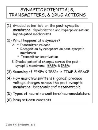
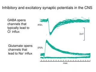
Inhibitory and excitatory synaptic potentials in the CNS. GABA opens channels that typically lead to Cl - influx. Glutamate opens channels that lead to Na + influx. Possible drug effects on synaptic effectiveness:. degradation of the neurotransmitter inside the axon terminal.

E N D
Inhibitory and excitatory synaptic potentials in the CNS GABA opens channels that typically lead to Cl- influx Glutamate opens channels that lead to Na+ influx
Possible drug effects on synaptic effectiveness: • degradation of the neurotransmitter inside the axon terminal. • increased neurotransmitter release into the synapse. • prevention of neurotransmitter release into the synapse. • inhibition of synthesis of the neurotransmitter. • reduced reuptake of the neurotransmitter from the synapse. • reduced degradation of the neurotransmitter in the synapse. • agonists (evoke same response as neurotransmitter) or antagonists (block response to neurotransmitter) can occupy the receptors. • enhance response of postsynaptic receptors to transmitter
PNS vs CNS CNS vs PNS
Neurons • a. About 1012 to 1014 of them in the primate central nervous system. • i. If you counted one neuron per second, it would take you approximately 1 million years to count all of these neurons. • ii. It is estimated that there are 5 X 10¹⁰ of them in the human cerebral cortex. • b. Most neurons have one of three roles based on their contribution to the general flow of information through the CNS: • i. Cell bodies of most sensory neurons lie outside of the CNS, and their processes extend into it. • ii. Cell bodies of motor neurons lie within the CNS, and their processes extend out of it. • Processing neurons lie entirely within the CNS and are called interneurons. • Nerves and ganglia together form the peripheral nervous system. • 1. Collections of processes from neurons = nerves if they areoutside the central nervous system and tracts if they are inside the central nervous system. • 2. Collections of nerve cell bodies outside the brain and spinal cord = peripheral ganglia. • a. For example, the cell bodies of many sensory neurons lie in a chain of ganglia that run parallel to most of the spinal cord.
A. Sensory (afferent) neurons form the input branch of the nervous system. • 1. In general, the neuronal cell bodies lie outside the CNS, e.g., the cell bodies of neurons that receive touch, pain, and temperature sensations lie in the spinal ganglia (often referred to as the dorsal root ganglia). • 2. Processes extend out to the location of the receptive field, and into the CNS. They synapse with cells in their segment of spinal cord, in other segments of the spinal cord, or in the brain, or all three. • 3. Excitation of sensory neurons is caused by a particular stimulus, usually involving some transfer of energy from the outer world to the neuron. • a. Particular sub-types of sensory neurons are specialized to receive particular types of stimulation. E.g., photoreceptors respond to light energy. • b. Sensory neurons respond to external stimulation by depolarizing; this process is called sensory transduction, because one form of energy - light, heat, mechanical movement, etc. - is changed into another form of energy - the electrical signal of the neuron. • c. In response to a stimulus, the receptor region of a sensory neuron depolarizes in a graded fashion. • i. The stronger the stimulus, the more they depolarize. This response is in some ways similar to a synaptic potential and is called a receptor potential. • ii. Most receptor potentials depend upon voltage-insensitive Na+ channels. • d. If the depolarization passes to the spike-initiating zone and exceeds its threshold, one or more action potentials travel from the spike-initiating zone to the CNS. The number of spikes and their frequency encode the intensity of a particular stimulus. • e. Each sensory neuron synapses with many neurons within the CNS. Multiple synapses produce divergence in neuronal circuitry. In most cases, the post-synaptic cells are interneurons. • f. The connections and the threshold properties of sensory neurons begin the work of processing signals from the outside world. Flow of information through the nervous system I
B. Interneurons continue the processing. • 1. Usually many neurons - sensory neurons and other interneurons - make synapses upon each central neuron = convergence. Convergence allows interneurons to serve an integrative function, integrating information from several input channels • 2. In general, synapses are made onto the dendrites and the soma. Axo-axonal synapses are rare. Hence in vivo, information flow through a single neuron is usually directed, from the dendrites, past or through the soma, and out the axon. • 3. Summation of the excitatory and inhibitory PSPs in an interneuron determines its activity level. • 4. There may be many levels of processing through a network of interneurons connecting sensory input to motor output. • 5. Much of the interesting work of the CNS - memory, "thinking," creativity - resides in networks of interneurons, most of which are located in the cerebral cortex. • C. Motor neurons are the efferent, or output, section of the nervous system. • 1. Dendrites and cell bodies of motor neurons lie within the CNS. Most of them are in the ventral portion of the spinal cord. Their axons extend into the periphery and end upon effector organs, particularly striated muscle fibers. • 2. These neurons direct "voluntary" movement and reflex movements. Flow of information through the nervous system II
Anatomy: Gray and White Matter • Gray matter • Unmyelinated nerve cell bodies • Dendrites • Axon terminals • White matter • Myelinated axons • Contains very few cell bodies
Anatomy: Bone and Connective Tissue • Brain is encased in bony skull or cranium • Spinal cord runs through vertebral column • Meninges lies between bone and tissues • Dura mater • Arachnoid membrane • Pia mater • Brain and spinal cord bathed in cerebrospinal fluid (CSF)
Anatomy: Blood-Brain Barrier Figure 9-6b
Neural Tissue: Metabolic Needs • Oxygen • Passes freely across blood-brain barrier • Brain receives 15% of blood pumped by heart • Glucose • Brain responsible for about half of body’s glucose consumption • Membrane transporters move glucose from plasma into the brain interstitial fluid • Hypoglycemia leads to confusion, unconsciousness, and death
Spinal Cord: Overview Central nervous system, posterior view Figure 9-4a
Spinal Cord: Anatomy Specialization in the spinal cord Figure 9-7a
Spinal Cord: Anatomy Figure 9-7b
Spinal Cord: Anatomy Propriospinal tracts remain within the cord Figure 9-7c (1 of 2)
Spinal Cord: Anatomy Figure 9-7c (2 of 2)
Spinal Cord: Integrating Center Figure 9-8
Geek Halloween costume “Dermatome Man” Spinal Cord: Dermatomes
Anatomy of the Brain Figure 9-9d
Cerebellum vs cerebrum Lateral view of brain The cerebellum coordinates movement Figure 9-9b
The Brain: Diencephalon Figure 9-10
Mid-Sagittal View of Brain Figure 9-9c
Gray Matter of the Cerebrum Figure 9-11
The Brain: The Limbic System • Emotion, memory, and learning Figure 9-13
Higher brain function Phrenology. In the 19th century, there was a pseudoscience (false science) called phrenology which claimed that different human traits were associated with bigger areas of the brain. The "science" of phrenology was created by Franz Joseph Gall and spread from Europe to the United States. By the later decades of the 1800s, there were popular phrenologists who were well paid to read the skulls of people and determine their strengths and weaknesses.
Brain Function: Cerebral Cortex • Three specializations • Sensory areas • Sensory input translated into perception • Motor areas • Direct skeletal muscle movement • Association areas • Integrate information from sensory and motor areas • Can direct voluntary behaviors
Mapping brain regions Wilder Penfield, 1940’s Electrical stimulation of exposed cortex during surgery
Brain Function: Functional Areas of the Cerebral Cortex Figure 9-15
Brain Function: Sensory Information • Special senses have devoted regions • Visual cortex • Auditory cortex • Olfactory cortex • Gustatory cortex • Processed into perception
Brain Function: Cerebral Lateralization Each lobe has special functions Figure 9-16