Download

1 / 86
860 likes | 1.06k Views
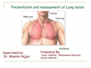
Presentation and management of Lung lesion. Supervised by: Dr. Wassim Hajjar. Presented By: Yaser Alshhab Mohammed Alkarzae Sultan Aldrees. Objectives:. To identify lung lesion and how to manage. To differentiate between Benign lung lesions.

E N D
Presentation and management of Lung lesion Supervised by: Dr. Wassim Hajjar Presented By: YaserAlshhab Mohammed Alkarzae Sultan Aldrees
Objectives: To identify lung lesion and how to manage. To differentiate between Benign lung lesions. Understand Solitary lung lesion and how to approach. Identify and treat Pulmonary carcinoid tumor. To have a clear picture about lung cancer. SCLC vs NSCLC. The surgical role in treating lung lesion.
Lung Lesion A lung lesion is abnormal tissue found on or in a person’s lung. It appear as lump of mass of tissues and other matter such as hardened blood, or pus in the lungs. They may appear in the bronchi or in the air sacs. Wherever they are found, they can cause pronounced difficulty in the respiratory system especially if they are malignant.
Tuberculosis creates cavities visible in x-rays like this one in the patient's right upper lobe. A CXR in a patient with central cancer of the right lung. Notice the white mass in the middle portion of the right lung In some cases, lung lesions may be numerous and may form on a specific area which is known as solitary lung lesion. In general, lung lesions may be classified as benign or malignant. Benign as : Malignant lung lesions: granulomateous lesion (TB), cyst carciniod lung tumor, benign tumors (hamartoma, Lung cancer(SCLC,NSCLC) bronchial adenoma, mucous gland adenoma)
Benign Lung Lesion Benign lung tumors are a heterogenous group of neoplastic lesions originating from pulmonary structures. These tumors include bronchial adenomas, hamartomas, and a group of uncommon neoplasms (eg, chondromas, fibromas, lipomas, leiomyomas, hemangiomas, teratomas) Although benign lung tumors do not pose a significant health problem, complications can result from an obstructive lesion that could predispose the patient to pneumonia, atelectasis, and hemoptysis. Age range is 17-77 years. The exact cause is unknown, because it is usually asymptomatic and discovered incidentally.
It is clinically asymptomatic . It may present with atelectesis ,airway obstruction <<suggesting an endobronchial origin. Majority identified incidentally finding on CXR. CT scanning and FNA occasionally suggest a benign diagnosis Definitive diagnosis is made by excisonal biopsy Rigid bronchoscopy diagnostic and therapeutic in case of symptomatic lesions
Hamartomas(chondroadenomas) Parenchymal hamartoma of the lung. The surrounding lung falls away from the well-circumscribed mass, a typical feature of these lesions. The hamartoma shows a variegated yellow and white appearance, which corresponds respectively to fat and cartilage Most common type of benign lung tumor, slow growing lesion Mainly occur in adults. Hamartomas are peripherally located. Grossly, they have a firm marblelike consistency. Histologically, hamartomas generally consist of epithelial tissue and other tissues such as fat and cartilage. Radiologically appears as well-circumscribed single nodules, up to 2cm , it can be localized in the lung CT scanning shows calcification and fat in up to half of the lesions.
Bronchial Adenomas Make up 50% of all benign pulmonary tumors. The use of the term bronchial adenoma should be discouraged because it encompasses several benign and malignant tumors. Mucous Gland Adenomas
Solitary lung lesion(Coin lesion) Definition: Is a peripheral circumscribed mass in the lung less than 3 centimeters in diameter. It can be an incidental finding found in up to 0.2% of chest X-raysand around 1% of CT scans The nodule most commonly represents a benign tumor such as a granuloma or hamartoma, but in around 10-20% of cases it represents a malignant cancer, (But smoker patients older than 50 years have >50% risk for lung cancer). Usually asymptomatic but it may include {coughing,hemoptysis,chest pain, and weight loss}
Chest X-ray showing a solitary pulmonary nodule (indicated by a black box) in the left upper lobe.
Pathophysiology A solitary pulmonary nodule is defined as a single, discrete pulmonary opacity that is less than 3 cm in diameter, surrounded by normal lung tissue, and not associated with adenopathy or atelectasis. Lesions larger than 3 cm are considered masses and are treated as malignancies until proven otherwise.Generally, a pulmonary nodule must reach 1 cm in diameter before it can be identified on a chest radiograph.
When it is significant:In the presence of risk factors for malignancy:
Approach We start by order the following investigation: 1- chest x ray 2-TB skin test 3-sputum cultures 4-sputum cytology(diagnostic in 5%-20%) 5-chest CT 6-biopsy In case of early bleeding we do bronchoscopy.
A CXR or chest CT scan can differentiate between malignant and benign lesion: 1) popcorn calcification Hamartoma 2) Concentric calcification calcified granuloma. 3)Spiculated lesions, with scattered calcification Are highly suspicious of malignancy.
Management: Based on the results of exams and tests, persons with SPN can be divided into the following 3 groups: .
Carciniod Tumor Carcinoid tumors are a slow-growing cancer that can arise in several places throughout your body. Carcinoid tumors, which are one subset of tumors called neuroendocrine tumors APUD{amine-precursor uptake and decarboxylation},known as the usually appear in the gastrointestinal tract (appendix, stomach, small intestine, colon, rectum) and in the lungs. Metastasis to the Liver with GI symptoms.
Types of Carcinoid tumor Typical lung carcinoid Atypical lung carcinoid
Typical Carcionoid Tumor Typical carcinoid tumors of the lung represent: Most well differentiated and least biologically aggressive type of cardconoid tumor. Grow slowly and tend to metastasize infrequently (5%-15%) Consist of polygonal cells arranged in clusters.
Atypical Carcinoid Tumor Atypical carcinoid tumors of the lung represent: More aggressive histologic and clinical picture. Metastasize at a considerably higher rate than do typical carcinoid tumors(predominantly lymph node) reported in 50-70% and, therefore, carry a worse prognosis. Variable nuclear configuration and moderate mitotic activity.
Carcinoid Syndrome Carcinoid syndrome has been reported in association with very large bronchopulmonarycarcinoid tumors or in the presence of metastatic disease. It is noted much less frequently in association with carcinoids of pulmonary origin than those originating within the gastrointestinal tract.
Frequency The gastrointestinal tract is the most common area in which carcinoid tumors arise. Bronchopulmonarycarcinoid tumors are reported to represent about 10% of all carcinoid tumors. One to 6% of all lung tumors are carcinoid tumors.(10% are atypical) Carcinoid tumors occur in equal numbers of males and females. The average age is 40-50 years,. Atypical carcinoid tumors appear in older people than typical carcinoids do. 90% of tumors develop within a bronchus.(10% of tumors arise in a mainstem bronchus; rarely appear in the trachea. ) 10% of tumors are located in the pulmonary periphery..
Clinical presenatation About 25% are asymptomatic. In symptomatic patients, Symptoms of bronchial obstruction: persistent cough, hemoptysis, recurrent or obstructive pneumonitis,Wheezing, chest pain, and dyspnea. Symptoms of malignancy: Weight loss, fatigability, and generalized illness.
Investigation Bronchoscopic image shows a red nodule that obstructs the entrance to the lower lobe bronchus Chest xray:An abnormal finding on chest X-ray is present in about 75% of patients with a carcinoid lung tumor Bronchoscopy: It reveals round red-yellow-purpule mass covered by epithelium that protrudes into bronchial lumen. Carcinoid tumor is Red shiny and it’s very easy to bleed
CT scan: Some carcinoid lung tumors that are small or covered by other organs in the chest may not be seen on a chest X-ray MRI: Same as CT but it help in differentiating small tumors from adjacent blood vessels Radionuclide : 1)Octreotide scintigraphy 2) Iodine-131 meta-iodo-benzyl guanidine (MIBG) scintigraphy Biopsy:Even if a chest X-ray and/or CT scan shows a tumor, these exams cannot confirm whether the mass is a carcinoid lung tumor, a lung carcinoma, or a localized infection.
Chest radiograph (CXR) demonstrates complete collapse of the left lower lobe. The cause of collapse is not identified on the image,so we asked for a ct scan. CT scan shows a hyper attenuating nodule within the left main bronchus. This is a bronchial carcinoid tumor.And then we need to ask for biopsy to confirm the diagnosis.
Managment All pulmonary carcinoid tumors should be treated as malignancies by doing total surgical resection as long as no surgery contraindication exist.
Medical Therapy No medical therapy exists for the primary treatment of carcinoid tumor of the lung. Chemotherapeutic agents and radiation therapy have been used in the treatment of metastatic disease(Carcinoid Syndrome)
Surgical Therapy Surgical resection is the primary mode of therapy . We have different types of surgical treatment,but anatomic lobectomy is the most commonly performed procedure at present for resection of pulmonary carcinoid tumors.
Types of surgery Most tumors follow a benign course and are amenable to surgery. Surgical options range from radical resection (the tumor with a good margin of normal tissue is removed) to minimally invasive surgery. Different surgical options include the following: Sleeve resection: Section of the airway containing the tumor is removed. In this procedure, we have to remove one part of the main bronchus containing the tumor and anastmose the bifurcation with bronchus intermmedius, instead of pneumonectomy to preserve the other tumor free lobes) Segmental resection: Segment of the lung containing the tumor is removed. Wedge resection: Small wedge of the lung containing the tumor is removed.
Lobectomy: Lobe of the lung containing the tumor is removed. Pneumonectomy: Entire lung containing the tumor is removed. (Rarely) Endoscopic tumor ablation using laser: This technique involves removal of the tumor through the bronchoscope, using laser. It is reserved for treating bronchial obstruction caused by the tumor or reduction of the tumor mass prior to surgical resection. This procedure is rarely curative on its own. Endoscopoic tumor ablation using laser, is not curative we use only in palliative treatment
Follow Up After discharging from the hospital, we need to follow up the pt for 2-3 months in the first year (clinically and chest x ray), then every 6 months for the following years. Prevention Unlike most lung tumors, carcinoid lung tumors have not been associated with smoking, air pollution, or other chemical exposures. Therefore, there are no known ways to prevent carcinoid lung tumors.
Prognosis Carcinoid lung tumors generally have a better prognosis(mainly typical) than other forms of lung cancer. Typical carcinoid lung tumors: 5-year survival rate of 78%-95% 10-year survival rate of 77%-90%. Atypical carcinoid lung tumors : 5-year survival rate of 40%-60% 10-year survival rate of 31%-60%.
Definition : Lung cancer is a disease which consists of uncontrolled cell growth in tissues of the lung. The vast majority of primary lung cancers are carcinomas, derived from epithelial cells.
The incidence of lung cancer has increased significantly over the last three decades. The most common cancer in male and the second in female ( after a breast cancer) It is the 1st most common cause of cancer-related death in both men and women.
Causes: • Smoking: • responsible for 85% of all lung cancers . • Passive smoking account for 3 % • ( nitrosamines and polycyclic aromatic hydrocarbons) • Environmental: • Asbestos , silica ,chromium, cadmium
Dietary: • cholesterol and high fat diet increases the risk. • Fruit and vegetables decreases the risk • Pre- existing lung lesion : • COPD, IPF , TB or infarct, Viruses ( HPV , CMV) • Inheritence:AD • Molecular genetics.
Symptoms and signs: For a primary tumor: respiratory symptoms constitute more than 70% of the presentation Cough, dyspnea , unresolved pneumonia, wheeze, hymoptysis, pain ) For a regional spread: ( pleural effusion, chest wall pain, pericardial effusion , SVS, horner syndrome, horesness, dysphagea , phrenic nerve injury.
Distant met : adrenal, liver, bone, brain. Paraneoplastic syndrome: ACTH, ADH, GH, calcitonin , PTH. SCLC : ACTH( cushing’s synd), ADH (SIADH) Sqamous cell canrcinoma : PTH > hyper calcimia Cachexia (weight loss, fatigue, and loss of appetite
Diagnosis: • Performing a chest radiograph is the first step if a patient reports symptoms that may suggest lung cancer. Performing a chest radiograph is the first step if a patient reports symptoms that may suggest lung cancer.
CT scanning: Has prove useful for assessing the primary tumor in relation to size and proximity to adjacent structure and surgical approaches
It is only 50% accurate in diagnosing tumor invasion into structures Abdominal CT is mandatory ( adrenal and liver
MRI Has no advantage over CT , but better in visualazing tumor incasion of nearby structures.