Download
1 / 141
1.41k likes | 1.44k Views
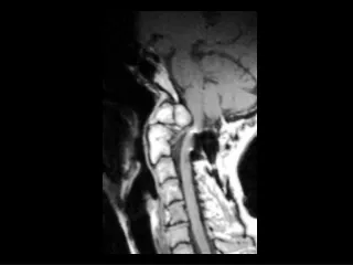
Os odontoidium. Findings: Complete separation of dens ossification center Posterior subluxation of the dens and the anterior arch of C1 Compression of the cervical cord ddx: Dens fracture. Choroid plexus papilloma of the 3 rd ventricle. Findings: Enhancing lobulated mass in the 3rd vent

E N D
Os odontoidium • Findings: • Complete separation of dens ossification center • Posterior subluxation of the dens and the anterior arch of C1 • Compression of the cervical cord • ddx: • Dens fracture
Choroid plexus papillomaof the 3rd ventricle • Findings: • Enhancing lobulated mass in the 3rd vent • Dilated ventricles • ddx: • Intraventricular meningioma • Metastasis
Carotid fibromuscular dysplasia • Findings: • irregular serial stenosis of ICA = “string of beads” • ddx: • NONE! • This is an Aunt Minnie!
Schwannoma • Findings: • Single enhancing extramedullary intradural lesion • ddx: • Meningioma • Drop metastasis
Cholesteatoma • Findings: • soft tissue mass in the left middle ear involving Prussak's space • erosion of scutum and ossicles • ddx: • NONE! • This is an Aunt Minnie!
Chondrosarcomaof the cricoid • Findings: • soft tissue mass involving right cricoid cartilage containing fine stippled calcifications • mild effacement of posterolateral airway • ddx: • Locally aggressive SCC • lymphoma • metastasis
Neurofibromatosis type I • Findings: • Numerous round and tubular isointense T1 lesions growing through the neural foramina • ddx: • NONE! • This is an Aunt Minnie!
CPA lipoma • Findings: • Large mass in the left cerebellopontine angle cistern • Hyperintense T1 signal • Fat attn on CT • ddx: (CPA mass) • CN 7/8 schwannoma • meningioma • glioma • metastasis • epidermoid • arachnoid cyst • AICA aneurysm
Dural sarcoid • Findings: • thick enhancing dura along rt frontoparietal convexity • associated parenchymal edema • meningeal enhancement • ddx: • meningioma • TB • metastasis
Foix-Alajouanine syndrome • Findings: • diffuse hyperintense T2 intramedullary cord signal • serpentine flow voids along dorsal surface of cord and enlarged draining veins on angiogram • venous hypertension and progressive myelopahty due to type I dural AVM • ddx: • MS • transverse myelitis • Tumor • Trauma • ischemia/infarction
Ganglioglioma • Findings: • faint hypointense FLAIR signal in the right medial temporal lobe without significant mass effect or edema • bright gyriform enhancement • ddx: • DNET • gangliocytoma • oligodendroglioma • focal encephalitis (if for edema)
Germinoma • Findings: • enhancing surprasellar mass causing hydrocephalus • dense on I- CT • ddx: • craniopharyngioma • lymphoma • meningioma
Disc herniation • Findings: • “paint brush” cut off of contrast column at the level of the disc space • ddx: • Other extradural lesions: • Hematoma • Discitis / abscess • Metastasis
Glomus jugulotympanicum • Findings: • enhancing soft tissue mass in the posterior left middle ear • involves jugular foramen, abuts carotid canal, and extends into EAC • stippled internal hypointense signal = flow voids • ddx: • NONE! • This is an Aunt Minnie!
Hypertensive encephalopathy • Findings: • T2 hyperintensity in bilateral frontal and parieto-occiptial lobes • Involvement of the coretx and white matter • ddx: • cyclosporine toxicity • dural sinus thrombosis
Von-Hippel Lindau • Findings: • Multiple enhancing masses in the posterior fossa and spinal cord with an associated syrinx • check the abdomen! • ddx: • NONE! • This is an Aunt Minnie!
Inverting papilloma • Findings: • large expansile mass involving right maxiallary sinus • extends in the nasal cavity and ethmoids cuasing bowing deformity but no destruction • ddx: • antrochoanal polyp • mucocele
Lacrimal lymphoma • Findings: • enhancing left preseptal mass, adjacent of the lacrimal gland • ddx: • hemangioma • Lacrimal gland tumor • Langerhan's cell histiocytosis • Metastasis
Extradural spinal lipomatosis • Findings: • Lobular high T1 signal mass lesion surrounding and compressing the spinal cord • obliteration of CSF space • ddx: • NONE! • This is an Aunt Minnie!
Laminar necrosis • Findings: • Symmetric abnormal enhancement of cerebral cortex • white matter attenuation abnormally low • no significant mass effect • causes: • global • profound anoxia • hypoglycemia • status epilepticus • focal • territorial infarction • ddx: • NONE! • This is an Aunt Minnie!
Laryngocele • Findings: • fluid density mass at the level of the right false cords • thin enhancing rim • ddx: • layngopyocele • abscess • ? necrotic LN
Chordoma • Findings: • Large mass arising from the sacrum • High T2 and intermediate T1 signal • ddx: • Chondrosarcoma • Lymphoma • Metastasis
Mesial temporal sclerosis • Findings: • Decreased volume of and increased T2 signal in the bilateral hippocamal formations • Surrounding white matter thinning • ddx: • NONE! • This is an Aunt Minnie!
Mucormycosis • Findings: • subperiosteal fluid collection in the left orbit • thickening of left medial rectus muscle • focal defects in lamina papyracia • Adjacent eithmoid sinus disease • ddx: • bacterial • other fungal
Spinal arachnoid cyst • Findings: • Tubular lesion along the posterior extradural space • Follows CSF signal • ddx: • Hematoma • Abscess • Lymphoma