Download
1 / 76
840 likes | 1.25k Views
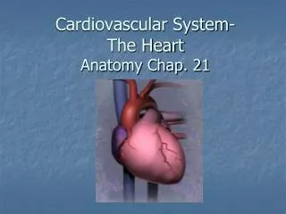
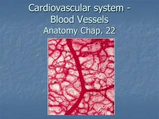
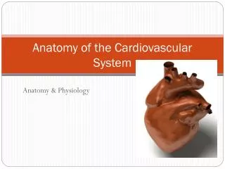
Anatomy of the Cardiovascular System. Cardiovascular System. As Also circulatory system Consists of: the heart, arteries, veins, capillaries. Heart. Four chamber muscular organ Comparable to the size of a closed fist Located in the mediastinum Behind sternum Between 2 nd and 6 th ribs

E N D
Cardiovascular System • As Also circulatory system • Consists of: the heart, arteries, veins, capillaries
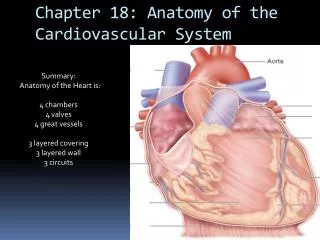
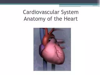
Heart • Four chamber muscular organ • Comparable to the size of a closed fist • Located in the mediastinum • Behind sternum • Between 2nd and 6th ribs • Between T5-T8 • Apex – base of heart • Located at the 5th intercostal space
Coverings of the Heart • Pericardium – loose fitting sac surrounding the heart • Fibrous pericardium – tough, loose-fitting, inelastic • Serous pericardium • Parietal layer: lines the inside of the fibrous pericardium • Visceral layer: adheres to outside of the heart • Pericardial space: between parietal and visceral layer • Filled with 10-15mL of pericardial fluid • Decreases friction
Walls of the Heart • Epicardium – outer layer • Epicardium = serous pericardium • Myocardium – thick, contractile layer composed of cardiac muscle cells • Intercalated disks contain many gap junctions • Allow cardiac muscle cells to function as a single unit syncytium • Endocaridium – interior of cardiac wall • Endothelial tissue • Covers projections of myocardial tissue called trabeculae
Chambers of the Heart • Atria – two superior chambers • “Receiving chambers” • Blood from veins enters atria • Ventricles – two inferior chambers • “pumping chambers” • Thick muscular walls to increase force of pumping action • Left > right • Separated by interventricular septum
Valves of the Heart • Permit blood flow in one direction during circulation • Atrioventricular valves (AV valves) • Also cuspid valves • Between atria and ventricles • Semilunar (SL valves) • Between R ventricle and pulmonary arteries and L ventricle and aorta
Atrioventricular Valves • Tricuspid valve • Btwn R atrium and ventricle • 3 flaps of endocardium • Connected to ventricular papillary muscle via chordae tendinae • Bicuspid valve • Btwn L atrium and ventricle • Also called mitral valve • Two flaps of endocardium
Semilunar Valves • Pulmonary semilunar valve • Btwn R ventricle and pulmonary trunk • Aorta semilunar valve • Btwn L ventricle and aorta
Chambers & Valves Trace the blood flow through the heart
Blood Supply to the Heart • Right and left coronary arteries • First branches off aorta • Right coronary artery right marginal artery & posterior interventricular artery • Left coronary artery circumflex artery & anterior interventricular artery • Most of the blood goes to the L ventricle • In 50% of the population, the R coronary artery is dominant
Blood Supply to the Heart • Anastomosis: Connections between blood vessels that allow for collateral circulation • Few exist between large branches of coronary arteries • In presence of an obstruction in a large artery ischemia will result to a large area of tissue • Myocardial infarction (MI) (aka heart attack) • Anastomoses do exists between smaller branches of the R and L coronary arteries
Blood Supply to the Heart • After traveling through the capillaries of the heart, blood empties into the R atrium via the coronary sinus
Conduction System of the Heart • Four structures composed of modified cardiac muscle • Sinoatrial Node (SA Node) • Pacemaker of the heart • 100s of cells in the R atrium near the opening of the superior vena cava • Atrioventricular Node (AV Node) • Small mass of cardiac muscle tissue • Left lower border of R atrium
Conduction System of the Heart • Atrioventricular Bundle • Also Bundle of His • Bundle of specialized cardiac muscle fibers originating in the AV node • Branches into R and L branches eventually becoming Purkinje fibers • Extend into the walls of the ventricles and papillary muscles
Warming Up I • The apex of the heart rests on the: • True/False: The epicardium and the fibrous pericardium are the same structure. • What is the correct order of the layers of the heart from outside to inside? • The ____________ connect the cuspid valves to the papillary muscles. • What is an anastomosis? • What is also known as the pacemaker of the heart?
Types of Blood Velssse • Artery – carries oxygenated blood away from the heart • “distributors” • Arteriole: small artery • Precapillary sphincters: regulate the blood flow into capillaries
Types of Blood Vessels • Vein – carries unoxygenated blood towards the heart • Great ability to stretch (capacitance) • Function as reservoirs: blood pools in the valves then is pushed forward from the pumping pressure • Venules: small vein
Types of Blood Vessels • Capillaries – arterial system switches to venous system • “primary exchange vessels” • Transport materials to and from the cells • Speed of blood flow decreases to increase contact time • Microcirculation: blood flow between arterioles, capillaries and venules
Structure of Blood Vessels • Tunica adventitia - outermost layer • Fibrous connective tissue • Holds vessels open; prevents tearing of vessels walls during body movements • Larger in veins than arteries • Tunica media – middle layer • Smooth muscle and elastic CT • Helps vessels constrict and dilate • Larger in arteries
Structure of Blood Vessels • Tunica intima – innermost layer • Composed of endothelium • Semilunar valves present in veins • One cell thick in capillaries
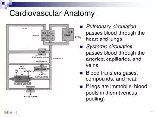
Circulatory Routes • Systemic Circulation – blood flow from the L ventricle to the body & back to the R atrium • Pulmonary Circulation – blood flow from the R ventricle to the lungs and back to the L atrium
Arch of aorta Subclavian (L and R) Brachiocephalic common carotid (L and R) Axillary (L and R) Brachial (L and R) Radial Ulnar Abdominal aorta Common iliac External iliac Femoral Popliteal Posterior tibial Anterior tibial Dorsal pedis Systemic Arteries
Superior vena cava Inferior vena cava External jugular Internal jugular Brachiocephalic (L and R) Subclavian (L and R) Cephalic axillary Basilic Median basilic Median cubital Common iliac External iliac Femoral Popliteal Great saphenous Small saphenous Systemic Veins
Fetal Circulation • Two umbilical arteries carry blood to the placenta • The placenta allows for exchange of oxygen and nutrients from the mother. Maternal and fetal blood do NOT mix. • Umbilical vein returns oxygenated blood and enters fetus via the umbilicus • Foramen ovale – hole btwn the R and L atria • Allows for blood to bypass the R ventricle and pulmonary circulation
Fetal Circulation • Ductus arteriosus – small vessel connecting the pulmonary artery and the aorta • Allows for another bypass route from the lungs **Most of fetal blood is a mixture of oxygenated and deoxygenated blood**
Changes After Birth • Umbilical vein become round ligament • Umbilical arteries become umbilical ligaments • Foramen ovale closes after first few breaths • Full closure may take up to 9 months • Ductus arteriosus contracts as soon as respirations begin • Become fibrous cord
Warm Up II • What is the function of the precapillary sphincters? • List the layers of arteries and veins from superficial to deep. How do the layers differ in arteries vs veins? • What type of tissue are capillaries composed of? • What blood vessel has valves? • Differentiate between pulmonary and systemic circulation.
Warm Up III Announcements: • Warm ups due today • Chapter 18 quiz next Tuesday – will finish notes on Monday • Heart and Blood Vessel Practical next Wednesday Warm Up: • Name the arteries that branch off the aorta. • What are the 4 divisions of the aorta? • True/False: During fetal circulation fetal and maternal blood mix. • Explain the function of the umbilical cord during fetal circulation. • Describe the foramen ovale and the ductus arteriosus.
Warm Up IV Announcements: • Chapter 18 Quiz tomorrow • Practical Wednesday (sheep heart and blood vessels) Warm Up: • Respectively, the right and left atrioventricular (AV) valves are also referred to as: • From the L ventricle, blood flows into the: • The pulmonary veins carry blood to the: • The most abundant blood supply goes to the: • What type of blood vessel has valves? What type of valves are they?
Pericardium Disorders • Pericarditis – inflammation of the heart • Causes: trauma, viral or bacteria infection, tumor • Edema causes visceral and parietal layers to rub together = chest pain • Pus or blood build up in pericardial space • S/S • Pain with respirations or coughing, dyspnea, restlessness • Complications: Pericardial Effusion, Cardiac Tamponade • Treatment: • Antibiotics, pain meds, antiinflammatory meds, pericardiocentesis (Cardiac Tamponade)
Heart Valve Disorders • General Principles: • Congenital defect: decreased pumping efficiency • Incompetent valve leak: allows backflow into previous chamber • Stenosed valves: narrowed valve; slowing blood from out of chamber
Heart Valve Disorders • Mitral Valve Prolapse (MVP) • Flaps of mitral valve extend back into L atrium causes leaking • Mostly genetic basis • 1 in 20 people • S/S: most asymptomatic; chest pain, fatigue • Treatment: valvuloplasty
Heart Valve Disorders • Aortic Regurgitation • Blood leaks back into L ventricle during ejection into the aorta • Volume overload in L ventricle, hypertrophy, dilation of L ventricle • Complications: myocaridal ischemia • Treatment: valvuloplasty
Myocardium Disorders • Atherosclerosis • Type of arteriosclerosis • Lipids build up on the inside of vessel walls calcify vessels hard & brittle • Risk factors: cigarette smoking, high fat/cholesterol diet, hypertension
Myocardium Disorders • Myocardial Infarction • “Heart Attack” • Coronary thrombosis: clot • Coronary embolism: mobilized clot • Occlude coronary artery heart tissue deprived of oxygen cell death • S/S: • Angina pectoris – severe chest pain resulting from inadequate oxygen to myocardium • Treatment: Coronary Bypass Surgery • Veins are harvested from other areas of the body and used to bypass obstructions