Download
1 / 22
220 likes | 515 Views
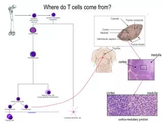
Where do T cells come from?. medulla. cortex. cortex. medulla. cortico-medullary junction. T cell development. Hematopoeitic Stem cell. Lymphoid progenitor. Lymphoblast. Prolymphocyte. Small lymphocyte. T lymphocyte. Lymphoblast. Lymphocyte. Development of the thymus.

E N D
Where do T cells come from? medulla cortex cortex medulla cortico-medullary junction
T cell development Hematopoeitic Stem cell Lymphoid progenitor Lymphoblast Prolymphocyte Small lymphocyte T lymphocyte Lymphoblast Lymphocyte
Development of the thymus The thymic epithelium develops as two flask-shape endodermal diverticula, arising from the third branchial pouch (pharyngeal pouch), and extending into the surrounding mesoderm and neural crest-derived mesenchyme in front of the ventral aorta. During the late stages of the development of the thymic epithelium, lymphoid cells from bone-marrow invade the thymus and aggregate to form lymphoid follicles. The thymus grows between birth and puberty and then begins to atrophy (hormone related). Proportional to thymic size, thymic activity (T cell output) is most active before puberty. Upon atrophy, the size and activity are dramatically reduced, and the organ is replaced primarily with fat (involution).
What happens if there is no thymus? “Nude” mice are defective and lack a functional thymus. As a result, they have virtually no T cells. The impact of this on the acquired immune response is profound: • No cell-mediated immune responses requiring CD4+ and/or CD8+ T cells • No killing of virus-infected or malignant cells (no cytotoxic T cells) • Defective antibody formation (no CD4+ helper T cells) • No graft rejection (requires both CD4+ and CD8+ T cells)
Double negative Double positive Single positive
T-cell development POSITIVE SELECTION Interaction with MHC class I or MHC class II on cortical epithelium NEGATIVE SELECTION Interaction with MHC class I or MHC class II + self peptide (dendritic cells, macrophages). AIRE protein expression (AutoImmune REgulator).
T cells interact with antigen presenting cells through the T cell receptor (TCR) and MHC T cell receptor CD4 MHC classII Helper T cell MHC classI Cytotoxic T cell CD8 Antigen presenting cell
T cell receptor genes rearrange in the thymus Similar rearrangements occur at thea and d (the d locus is embedded in thealocus), b, and gloci. Thealocus is more diverse than theb(~100Vacompared to ~20 Vb). Theglocus is organised more similar to an antibodylight chain locus than the other TCR loci. Vb (~20) Jb1 (7) Db1 Jb2 (6) Vb1 Vb2 Vbn D J D J J J J J J Cb1 J J J J J J Vblocus (mouse) rearranged DNA D J Cb1 Vb1 spliced mRNA transcript J Cb1 D Vb1
What happens next when T cells leave the thymus? Lymphoid tissue homing receptors CD62L+, CCR7+ T cells B cell area T cell area blood vessel Homing to mucosae and sites of infection CD62L-, integrins a4b7+/a4b1+, CCR5+, CCR3+ T cells CD34 L-selectin T diapedesis T Naïve T cell LFA-1 High endothelial venule ICAM-1
T cell circulation through the lymph node Dendritic cell interaction primes T cell B cell area T cell area T cell medullary chords dendritic cell T cell afferent lymphatic blood vessel High Endothelial Venule (HEV) efferent lymphatic germinal centres T cells provide help for B cell expansion
T cell activation… complexity prevents inappropriate activation Dendritic cell CD80/86 CD40 MHC II CD28 TCR T CD40L IL-4 T cell B cell ICOS ICAM-1 (CD54) LFA-1 (CD11/CD18) CD80 LFA-3 (CD58) CD2 CD40L (CD154) CD40 CD4 peptide CD3 MHC II T-cell receptor CD4 (CD80) CD28 Only professional APCs express the gallimaufry of co-stimulatory molecules required to activate naïve T cells! CD28 (CD86) T cell Dendritic cell
There are several functional subsets of CD4+ T cells Not all pathogens are the same, therefore the immune system has evolved ways of tailoring the immune response to suit the pathogen. Simplistically, Th1 cells promote a cellular response. Type I pathogens (bacteria/viruses) salmonella HIV NK cell IL-12 mDC1 Antigen, IL-12 + IL-18 IFN-g IFN-g IL-18Ra IL-12Rb2 Th1 LT-a IL-12Rb1/b2 IL-12Rb1 IL-18R IL-12R T cell • Cellular Immunity • Tc cell proliferation • Increase macrophage activity • Help B cells produce antibody IL-12
There are several functional subsets of CD4+ T cells Th2 cells produce cytokines that favour a humoral response and induce mast cell production. Type 2 pathogens (helminths) Ancylostoma caninum IL-12Rb1 IL-12Rb1 mDC2 IL-4 IL-5 IL-13 IL-25 IL-12Rb2 Th2 IL-4 Antigen IL-5 Basophil Eosinophil Mast cell • Humoral Immunity • B cell proliferation • Class switching e.g. IgA & IgE • Increase Ig production IL-25 c-kit+ NKT Immunity to parasites
There are several functional subsets of CD4+ T cells Th17 cells produce cytokines that promote neutrophil responses. iDC Treg TGF-b Antigen, IL-23 + IL-1/18 IL-6 mDC IL-17A IL-17F IL-6 G-CSF IL-23R Th17 IL-12Rb1 IL-23R IL-12Rb1 IL-18R IL-23R IL-23 Type 17 pathogens (bacteria, fungi) G-CSF Enhance neutrophil response aspergillus salmonella
Cytotoxic T cells kill virus-infected cells cytotoxic T cell “cytotoxin”-containing granules apoptosis virus-infected cells Perforin – aids delivery of granule contents into cytoplasm of target cell Granzymes – Serine proteases, activate apoptosis by switching on Caspases Granule contents Granulysin – Activates apoptosis IFN-γ – Inhbits viral replication, increases MHC I. Macrophage activation TNF-α, – Macrophage activation, apoptosis induction. Cytokines
…and finally… NK cells NK cells recognise “stress” receptors on the surface of virus-infected and malignant cells. They can also detect reduced MHC expression. NK cell Infected cell NK cells can kill infected cells by ADCC, they are rich in Fc receptor expression. An inherited deficiency in NK cells has been described. Manifesting as recurrent EBV, CMV, VZV and HSV infections, NK cells must play an integral role in immunity to herpes viruses.
Summary - Lymphocytes and their function(s) Helper T cell CD4 Provides help to other cells of the immune system to respond to infection T cell receptor Cytotoxic T cell CD8 Kills cells infected with intracellular pathogens NK cell Innate immunity - cell killing and cytokine secretion