Chapter #7
Chapter #7. Skeletal System. Chapter 7.1 Introduction. Bones the organs of the skeletal system Provide points of attachment for muscles Protect soft tissue/organs Support soft tissue House blood-producing cells Store inorganic salts Provide passageways for blood vessels and nerves

Chapter #7
E N D
Presentation Transcript
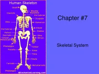
Chapter #7 Skeletal System
Chapter 7.1 Introduction • Bones the organs of the skeletal system • Provide points of attachment for muscles • Protect soft tissue/organs • Support soft tissue • House blood-producingcells • Store inorganic salts • Provide passageways for blood vessels and nerves • There are 206 bones in the adult human body
Chapter 7.2 Bone Structure • Epiphysis (epifisis) end of the bone • Articular cartilage covers end of bones • Diaphysis (diafisis) the shaft of the bone
Periosteum is a tough vascular covering of fibrous tissue. Bleeds when you fracture your bones.
Compact bone Spongy bone • Compact bone is a type of bone with a continuous matrix with no gaps. (Diaphysis) • Spongy bone is a type of bone with numerous branching bony plates. (Epiphysis)
Medullary cavity the hollow chamber/tube in long bones. Endosteum lines the medullary cavity. Lies under the periosteum. Marrow is a specialized type of soft connective tissue that fills the medullary cavity.
Osteocytes bone cells. • Lacunae very small, bony chambers. • Haversian canals (Central canals) • Osteon cylinder-shaped unit. • Volkmann’s canal perforating canals
Chapter 7.3 Bone Development and Growth • Intramembranous bones originate between sheetlike layers of connective tissue. • Osteoblasts bone-forming cells.
Endochondral bones-Long Bones they develop from masses of cartilage shaped like future boney structures.
Epiphyseal plate is the portion of bone where growth happens. Bones continue to grow until the plate closes. • If an Epiphyseal plate is damaged before it ossifies, elongation of the long bone may cease prematurely, or growth maybe uneven.
Chapter 7.4 Bone Function • Hemopoiesis the process of blood cell formation. Begins in the yolk sac, which lies outside the human embryo. • Marrow is a soft, netlike mass of connective tissue. • Red Marrow- makes red blood cells, white blood cells, and platelets. It is red because it contains hemoglobin. It occupies the cavities of most bones in an infant. In an adult, RM is found in the spongy bone of the skull, ribs, sternum, clavicles, vertebrae, and pelvis. • Yellow marrow- stores fat and is inactive in blood cell production.
Bone tissue is rich in calcium phosphate. • When blood is low in Ca, the body makes osteoclasts (breakdown bone) and release calcium into the blood for the body to use for metabolic processes. • Osteoporosis- lose of bone volume and mineral content. Factors that increase osteoporosis are low calcium intake, lack of physical exercise, decrease in blood estrogen.
Chapter 7.5 Skeletal Organization • Axial Skeleton • Skull, Ribs, Vertebral column • Appendicular skeleton • Arms, legs, and bones that anchor the limbs to the axial skeleton.
Chapter 7.6 Skull • Fontanels (Soft Spots)- membranous areas. Found in infants skulls they will close as they develop. Infant skull
Chapter 7.7 Vertebral Column • Atlas- 1st cervical vertebrae • Axis- 2nd cervical vertebrae
Chapter 7.8 Thoracic Cage • Thoracic cage includes ribs, thoracic vertebrae and sternum.
Chapter 7.9 Pectoral Girdle • Pectoral girdle (Shoulder girdle) includes the clavicles and scapulae.
Chapter 7.10 Upper Limb • Know the bone identification sheet
7.11 Pelvic Girdle • Includes two hipbones (coxae) • Ilium (hip bones) • Ischium (butt bones) • Pubis (where the 2 hip bones meet) Male Female
Chapter 7.12 Lower Limb • Know bone identification sheet
Chapter 7.13 Joints • Joints are functional junctions between bones. • Fibrous joints • Cartilaginous joints • Synovial joint • Ball-and-socket Joints • Condyloid • Gliding • Hinge • Pivot • Saddle
Fibrous joints These joints are also called "fixed" or "immoveable" joints, because they do not move. These joints have no joint cavity and are connected via fibrous connective tissue. The skull bones are connected by fibrous joints.
Cartilaginous joints These joints also have no joint cavity and the bones are connected tightly to each other with cartilage. These joints only allow a small amount of movement, so are also called "partly" or "slightly moveable" joints. The vertebrae are examples of cartilaginous joints.
Most of the joints in the body are synovial joints. These joints are "freely moveable" and are characterised by being surrounded by an articular capsule which contains the synovial fluid. Synovial fluid lubricates the joints, supplies nutrients to the cartilage and it contains cells that remove microbes and debris within the joint cavity. Because of the larger range of movements of these joints, there is an increased risk of injury example dislocations. Synovial joints are located predominantly in limbs. • Many synovial joints also have ligaments either inside or outside the capsule.
Synovial Joints • Ball and socket - allows movement around 3 axes - flexion / extension, abduction / adduction and rotation, ex. shoulder, hip. • Condyloid– permits a variety of movements. Examples joint between the metacarpals and phalanges. • Gliding - Flat bone surfaces allow side to side and backwards and forwards movements ex. between carpals, tarsals, between the sternum and the clavicle (sterno-clavicular) and the scapula and the clavicle. • Hinge - movement occurs primarily in a single plane ex. elbow, knee , ankle, interphalangeal joints. • Pivot- a ring of bone and ligament surrounds the surface of the other bone - movement in one plane, primarily rotation ex. between the atlas and axis (ie the cervical vertebrae numbers 1 and 2) and the radius and ulna. • Saddle joints - example thumb.
Work Cited • “Femur”. March 6, 2007. http://webschoolsolutions.com/patts/systems/bone-growth.gif • “Aged femur” and “Long bone”. March 6, 2007. http://images.google.com/imgres?imgurl=http://www.curehandpain.com/images/bone/spongy_bone.gif&imgrefurl=http://www.curehandpain.com/pages/bone/bone.php&h=226&w=347&sz=23&hl=en&start=40&tbnid=ZzjWovrGxZeNyM:&tbnh=78&tbnw=120&prev=/images%3Fq%3Dcompact%2Bbone%26start%3D20%26gbv%3D2%26ndsp%3D20%26svnum%3D10%26hl%3Den%26sa%3DN • “Intramembranous bones”. March 6, 2007. http://academic.wsc.edu/faculty/jatodd1/351/intramembranous_bone_growth.jpe • “Bone development”. March 6, 2007. http://classes.aces.uiuc.edu/AnSci312/Bone/Gil%209%2014%20Endochondral%20ossification.jpg • “X-ray”. March 6, 2007. http://www.shoppingtrolley.net/images/epiphyseal-plate.gif • “Plate injury”. March 6, 2007. http://cal.vet.upenn.edu/saortho/chapter_34/34F4.jpg • “Pelvis, lower limb and upper limb”. March 7, 2007. http://www.gpc.edu/~jaliff/spinchar.gif • “Vertebral Column”. March 7, 2007. http://www.shockfamily.net/skeleton/SPINE.JPG • “Rib Cage”. March 7, 2007. http://www.netterimages.com/images/vpv/000/000/007/7191-0550x0350.jpg • “Skull”. March 7, 2007. http://upload.wikimedia.org/wikipedia/commons/thumb/5/53/Gray_188_-_Side_view_of_the_skull.png/180px-Gray_188_-_Side_view_of_the_skull.png • “Infant Skull”. March 7, 2007. http://faculty.clintoncc.suny.edu/faculty/Michael.Gregory/files/Bio%20102/Bio%20102%20lectures/Motor%20Systems/infant_skull.jpg • “Skull joints”. March 9, 2007. http://faculty.clintoncc.suny.edu/faculty/Michael.Gregory/files/Bio%20102/Bio%20102%20lectures/Motor%20Systems/immovable_joint.jpg • “Vertebrae joint and synovial joint”. March 9, 2007. http://www.shoppingtrolley.net/images/anatomy/cartilaginous-joint.jpg