Sight
Sight. Visual Accessory Organs eyelids lacrimal apparatus extrinsic eye muscles. Eyelid. palpebra composed of four layers skin muscle connective tissue conjunctiva orbicularis oculi - closes levator palperbrae superioris – opens tarsal glands – secrete oil onto eyelashes

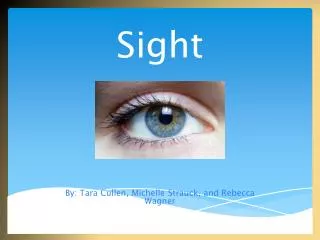
Sight
E N D
Presentation Transcript
Sight • Visual Accessory Organs • eyelids • lacrimal apparatus • extrinsic eye muscles
Eyelid • palpebra • composed of four layers • skin • muscle • connective tissue • conjunctiva • orbicularis oculi - closes • levator palperbrae superioris – opens • tarsal glands – secrete oil onto eyelashes • conjunctiva – mucous membrane; lines eyelid and covers portion of eyeball
Lacrimal Apparatus • lacrimal gland • lateral to eye • secretes tears • canaliculi • collect tears • lacrimal sac • collects from canaliculi • nasolacrimal duct • collects from lacrimal sac • empties tears into nasal cavity
Extrinsic Eye Muscles • Superior rectus • rotates eye up and medially • Inferior rectus • rotates eye down and medially • Medial rectus • rotates eye medially
Extrinsic Eye Muscles • Lateral rectus • rotates eye laterally • Superior oblique • rotates eye down and laterally • Inferior oblique • rotates eye up and laterally
Structure of the Eye • hollow • spherical • wall has 3 layers • outer fibrous tunic • middle vascular tunic • inner nervous tunic
Outer Tunic • Cornea • anterior portion • transparent • light transmission • light refraction • Sclera • posterior portion • opaque • protection
Middle Tunic • Iris • anterior portion • pigmented • controls light intensity • Ciliary body • anterior portion • pigmented • holds lens • moves lens for focusing • Choroid coat • provides blood supply • pigments absorb extra light
Anterior Portion of Eye • filled with aqueous humor
Lens • transparent • biconvex • lies behind iris • largely composed of lens fibers • elastic • held in place by suspensory ligaments of ciliary body
Ciliary Body • forms internal ring around front of eye • ciliary processes – radiating folds • ciliary muscles – contract and relax to move lens
Accommodation • changing of lens shape to view objects
Iris • composed of connective tissue and smooth muscle • pupil is hole in iris • dim light stimulates radial muscles and pupil dilates • bright light stimulates circular muscles and pupil constricts
Aqueous Humor • fluid in anterior cavity of eye • secreted by epithelium on inner surface of the ciliary body • provides nutrients • maintains shape of anterior portion of eye • leaves cavity through canal of Schlemm
Inner Tunic • retina • contains visual receptors • continuous with optic nerve • ends just behind margin of the ciliary body • composed of several layers • macula lutea – yellowish spot in retina • fovea centralis – center of macula lutea; produces sharpest vision (you move your eye to put images here) • Only cones (no rods) • optic disc – blind spot; contains no visual receptors • vitreous humor – thick gel that holds retina flat against choroid coat
Posterior Cavity • contains vitreous humor – thick gel that holds retina flat against choroid coat
Vision • Pass through: • Cornea • Aqueous humor • Lens • Vitreous humor • Retinal layers • Photoreceptor cells
Major Groups of Retinal Neurons • receptor cells, bipolar cells, and ganglion cells - provide pathway for impulses triggered by photoreceptors to reach the optic nerve • horizontal cells and amacrine cells – modify impulses
Light Refraction • Refraction • bending of light • occurs when light waves pass at an oblique angle into mediums of different densities
Types of Lenses Convex lenses cause light waves to converge Concave lenses cause light waves to diverge
Focusing On Retina • as light enters eye, it is refracted by • convex surface of cornea • convex surface of lens • image focused on retina is upside down and reversed from left to right
Visual Receptors • Rods • long, thin projections • contain light sensitive pigment called rhodopsin • hundred times more sensitive to light than cones • provide vision in dim light • produce colorless vision • produce outlines of objects • Cones • short, blunt projections • contain light sensitive pigments called erythrolabe, chlorolabe, and cyanolabe • provide vision in bright light • produce sharp images • produce color vision
Visual Pigments • Rhodopsin • light-sensitive pigment in rods • decomposes in presence of light • triggers a complex series of reactions that initiate nerve impulses • impulses travel along optic nerve • Pigments on Cones • each set contains different light-sensitive pigment • each set is sensitive to different wavelengths • color perceived depends on which sets of cones are stimulated • erythrolabe – responds to red • chlorolabe – responds to green • cyanolabe – responds to blue
Stereoscopic Vision • provides perception of distance and depth • results from formation of two slightly different retinal images
Life-Span Changes • Age related hearing loss due to • damage of hair cells in organ of Corti • degeneration of nerve pathways to the brain • tinnitus • Age-related visual problems include • dry eyes • floaters (crystals in vitreous humor) • loss of elasticity of lens • glaucoma • cataracts • macular degeneration
Clinical Application Refraction Disorders • concave lens corrects nearsightedness (myopia) • convex lens corrects farsightedness (hyperopia)