Download
1 / 107
1.08k likes | 1.34k Views
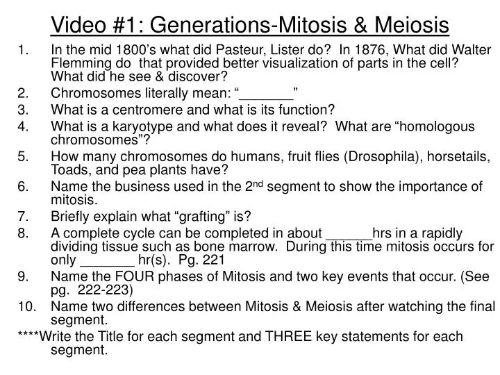
Video #1: Generations-Mitosis & Meiosis. In the mid 1800’s what did Pasteur, Lister do? In 1876, What did Walter Flemming do that provided better visualization of parts in the cell? What did he see & discover? Chromosomes literally mean: “_______”

E N D
Video #1: Generations-Mitosis & Meiosis • In the mid 1800’s what did Pasteur, Lister do? In 1876, What did Walter Flemming do that provided better visualization of parts in the cell? What did he see & discover? • Chromosomes literally mean: “_______” • What is a centromere and what is its function? • What is a karyotype and what does it reveal? What are “homologous chromosomes”? • How many chromosomes do humans, fruit flies (Drosophila), horsetails, Toads, and pea plants have? • Name the business used in the 2nd segment to show the importance of mitosis. • Briefly explain what “grafting” is? • A complete cycle can be completed in about ______hrs in a rapidly dividing tissue such as bone marrow. During this time mitosis occurs for only _______ hr(s). Pg. 221 • Name the FOUR phases of Mitosis and two key events that occur. (See pg. 222-223) • Name two differences between Mitosis & Meiosis after watching the final segment. ****Write the Title for each segment and THREE key statements for each segment.
Introductory Questions #1 1) How much DNA does a typical human cell have? How are chromosomes differ from chromatin? 2) How is a somatic cell different from a gamete? 3) How is every species different in regards to their chromosomes? 4) Name the main stages of the cell cycle. (pg. 221) 5) What are the four stages of mitosis? Which stage is the longest and which stage is the shortest? 6) Give three specific events that occur during prophase. How is Prometaphase different from prophase? 7) How are plant cell different from animal cells when they divide?
Introductory Questions #2 • What are the three checkpoints of the cell cycle that regulates mitosis? Which one is considered the “restriction point”? Why this checkpoint and not the others? • Name the two protein molecules that are high in concentration during the mitotic (M) phase of the cell cycle. Name the complex that it forms. • Why are telomeres considered to be a “mitotic clock”? DO telomeres contain genes? What does telomerase do? (see pgs 306-307 in Ch. 16) • How are tumor supressor genes different from an oncogenes? How is a proto-oncogene different from an oncogene? What kind of a protein does the RAS gene produce? • What is the difference between a malignant tumor and a benign tumor? • When looking at the hypothetical sequence of how mitosis may have evolved how is the process different in a bacteria and diatom from a plant and animal cell?
Introductory Questions #3 • How is a totipotent stem cell different from a pluripotent stem cell? See pgs. 415-418 (Ch. 21) • Which phase is used to obtain pictures of chromosomes in order to generate a karyotype • Give five differences between Mitosis and Meiosis. • Name three factors in Meiosis & reproduction that contributes in increasing genetic variability within a population. • What is a polar body? How is oogenesis different from spematogenesis? • How is a sporophyte different from a gametophyte? What do they produce and what process is involved, mitosis or meiosis? • What is a tetrad? Which phase of Meiosis does crossing over occur?
Mitosis and Meiosis • Chapter 12 & 13 • Mitosis & Meiosis
Next Unit: Genetics & DNA • Chapter 12 & 13: Mitosis & Meiosis • Chapter 14: Principles of Heredity • Chapter 15: Human Genetics & Disorders • **Two Labs will be done for this Unit • Goal: to complete before Thanksgiving and to take Test #3 on 11/25 (Tuesday)
Mitosis • Occurs only in certain types of cells • Form of asexual reproduction • Produces two genetically identical cells from one cell. • The splitting or dividing of the nucleus • Viewed in different stages by examining chromosome formation and behavior.
Significance of Understanding Mitosis • Preserves the continuity of life • Allows organisms to grow, repair, and reproduce • Important in unlocking the mysteries of embryonic development & stem cells • Important in understanding how cancer develops and could someday provide clues in stopping cancer.
Cell replacement (seen here in skin) Deadcells Epidermis, the outer layer of the skin Dividingcells Dermis Figure 8.11B
Packaging of Genetic Materialhttp://www.biostudio.com/demo_freeman_dna_coiling.htm Structure / ActivityDiameter • DNA: smallest structure about (2 nm) • DNA & Histones = Nucleosome (10 nm) • Chromatin Fibers** (30 nm) • Extensive Looping (300 nm) • Further Condensing (700 nm) • Fully Formed Chromosome (1400 nm)
Chromosomes • Condensed DNA attached to proteins • Can only be seen when a cell is actively undergoing mitosis. • Typical humans form 46 chromosomes vs. other organisms which varies significantly. • Our 46 chromosomes are thought to contain anywhere from 25,000 to 100,000 genes. • Duplicated before mitosis occurs producing a sister chromatid (identical copy) • Sister chromatids held together by “Centromere”
Cells from an onion Root tip • When the cell cycle operates normally, mitotic cell division functions in: • Growth (seen here in an onion root) Figure 8.11A
E. coli dividing Figure 8.3x
Asexual reproduction (seen here in a hydra) Figure 8.11C
THE EUKARYOTIC CELL CYCLE AND MITOSIS • A eukaryotic cell has many more genes than a prokaryotic cell • The genes are grouped into multiple chromosomes, found in the nucleus • The chromosomes of this plant cell are stained dark purple Figure 8.4A
Human male bands Figure 8.19x3
Human female karyotype Figure 8.19x2
Sister chromatids • Before a cell starts dividing, the chromosomes are duplicated Centromere • This process produces sister chromatids Figure 8.4B
When the cell divides, the sister chromatids separate Chromosomeduplication Sister chromatids Centromere • Two daughter cells are produced • Each has a complete and identical set of chromosomes Chromosomedistributiontodaughtercells Figure 8.4C
Interphase • Cells spend most of its time in this phase • Cells are growing • DNA has to be replicated (all 2 meters of it) • Proteins are being produced • 90% of all cells are in this phase • Three phases: G1, S, and G2
Prophase • Chromatin thickens (coils) into chromosomes • Two copies of DNA are present: sister chromatids (twice the amount of DNA is present) • Centrioles replicate forming another centrosome separate. • Centrioles separate to each side of the nucleus • Nuclear membrane (envelope) disappears • Microtubules elongate forming the spindle apparatus
Metaphase • Chromosomes align themselves up in the center of the cell • Spindle fibers (microtubules) attach to the centromere of the chromosomes • Longest phase of Mitosis
Mitotic spindle Figure 8.6x2
Anaphase • Chromosomes separate by the shortening of the microtubules. • The sister chromatids separate to each side (pole) of the cell. (humans: 46 to each side) • The centrosome is located at each side of the cell.
Cytokinesis: Plant vs Animal Cells • Cleavage furrow: animals cells • Cell plate: Plant cells
Cytokinesis differs for plant and animal cells • In animals, cytokinesis occurs by cleavage • This process pinches the cell apart Cleavagefurrow Cleavagefurrow Contracting ring ofmicrofilaments Figure 8.7A Daughter cells
Cell plateforming Wall ofparent cell Daughternucleus • In plants, a membranous cell plate splits the cell in two Cell wall New cell wall Vesicles containingcell wall material Cell plate Daughtercells Figure 8.7B
Cells from an onion Root tip • When the cell cycle operates normally, mitotic cell division functions in: • Growth (seen here in an onion root) Figure 8.11A
Sea urchin development Figure 8.0x
Cell cycle collage Figure 8.5x
Fibroblast growth Figure 8.8x
The Cell Cycle: Generation Time • Interphase: most of a cell’s life (90%) -G1: 1st gap of growth -S phase: DNA is duplicated (synthesized) -G2 phase: 2nd gap of growth • Mitosis: splitting of the nucleus (PMAT) • Cytokinesis: separation of the cytoplasm
The cell cycle multiplies cells • The cell cycle consists of two major phases: • Interphase, where chromosomes duplicate and cell parts are made • The mitotic phase, when cell division occurs Figure 8.5
See Pgs 222-223 INTERPHASE PROPHASE Prometaphase Figure 8.6
METAPHASE ANAPHASE TELOPHASE AND CYTOKINESIS Cleavagefurrow Nucleolusforming Metaphaseplate Nuclearenvelopeforming Spindle Daughterchromosomes http://highered.mcgraw-hill.com/sites/0072437316/student_view0/chapter11/animations.html#
The Kinetochores • Located in the middle of each sister chromatid. • Microtubules attach and breakdown as the sister chromatids are pulled to opposite sides of the cell. • See Research on Pg. 225
Mitosis collage, light micrographs Figure 8.6x1
Evolution of Mitosis (pg. 227) Chromosomes attach to the plasma membrane Chromosomes attach to the nuclear membrane Pass through the nucleus Spindle forms within the nucleus
Regulation of Cell Division • Driven by specific molecular signals • Research has shown: • Two cells in different phases causes the other to be pushed into the next phases. • Ex. • S phase & G1 grown together will cause the G1 cell to enter into the S phase immediately • M phase cell & G1 cell will cause the G1 cell to enter into the M phase immediately. • There is an obvious control system in place.