Trichinella Spiralis
160 likes | 1.06k Views
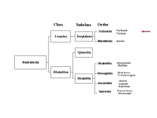
Trichinella Spiralis. INTRODUCTION. Trichinella is a nematode.

Trichinella Spiralis
E N D
Presentation Transcript
INTRODUCTION Trichinella is a nematode. Trichinellosis develops after the ingestion of meat containing cysts of Trichinella (e.g., pork or other meat from a carnivore). Although most infections are mild and asymptomatic, heavy infections can cause severe enteritis, periorbital edema, myositis, and (infrequently) death. (Harrison’s Principles of Internal Medicine , 17 Edition Volume 1, p.1316) (Harrison’s Principles of Internal Medicine , 18 Edition) T. spiralis, which is found in a great variety of carnivorous and omnivorous animal, is distributed world wide. (Harrison’s Principles of Internal Medicine , 17 Edition Volume 1, p.1316) (Harrison’s Principles of Internal Medicine , 18 Edition)
LIFE CYCLE After human consumption of trichinous meat, encysted larvae are liberated by digestive acid and proteases (Picture 1). The larvae invade the small-bowel mucosa and mature into adult worms. After 1 week, female worms release newborn larvae that migrate via the circulation to striated muscle. The larvae then encyst by inducing a radical transformation in the muscle cell architecture. Although host immune responses may help to expel intestinal adult worms, they have little effect on muscle-dwelling larvae. (Harrison’s Principles of Internal Medicine , 17 Edition Volume 1, p.1316) (Harrison’s Principles of Internal Medicine , 18 Edition)
EPIDEMIOLOGY Human trichinellosis is often caused by the ingestion of infected pork products and thus can occur in almost any location where the meat of domestic or wild swine is eaten. Human trichinellosis also may be acquired from the meat of other animals, including dogs (in parts of Asia and Africa), horses (in Italy and France), and bears and walruses (in northern regions). Although cattle (being herbivores) are not natural hosts of Trichinella, beef has been implicated in outbreaks when contaminated or adulterated with trichinous pork. Laws that prohibit the feeding of uncooked garbage to pigs have greatly reduced the transmission of trichinellosis in the United States. About 12 cases of trichinellosis are reported annually in this country, but most mild cases probably remain undiagnosed. Recent U.S. and Canadian outbreaks have been attributable to consumption of wild game (especially bear meat) and, less frequently, of pork. (Harrison’s Principles of Internal Medicine , 17 Edition Volume 1, p.1316) (Harrison’s Principles of Internal Medicine , 18 Edition)
PATHOGENESIS AND CLINICAL FEATURES Clinical symptoms of trichinellosis arise from the successive phases of parasite enteric invasion, larval migration, and muscle encystment (Picture 1). Most light infections (those with <10 larvae per gram of muscle) are asymptomatic, whereas heavy infections (which can involve >50 larvae per gram of muscle) can be life-threatening. Invasion of the gut by large numbers of parasites occasionally provokes diarrhea during the first week after infection. Abdominal pain, constipation, nausea, or vomiting also may be prominent. (Harrison’s Principles of Internal Medicine , 17 Edition Volume 1, p.1316) (Harrison’s Principles of Internal Medicine , 18 Edition) Symptoms due to larval migration and muscle invasion begin to appear in the second week after infection. The migrating Trichinella larvae provoke a marked local and systemic hypersensitivity reaction, with fever and hypereosinophilia. Periorbital and facial edema is common, as are hemorrhages in the subconjunctivae, retina, and nail beds ("splinter" hemorrhages). A maculopapular rash, headache, cough, dyspnea, or dysphagia sometimes develops. Myocarditis with tachyarrhythmias or heart failure—and, less commonly, encephalitis or pneumonitis—may develop and accounts for most deaths of patients with trichinellosis. (Harrison’s Principles of Internal Medicine , 17 Edition Volume 1, p.1316) (Harrison’s Principles of Internal Medicine , 18 Edition)
Upon onset of larval encystment in muscle 2–3 weeks after infection, symptoms of myositis with myalgias, muscle edema, and weakness develop, usually overlapping with the inflammatory reactions to migrating larvae. The most commonly involved muscle groups include the extraocular muscles; the biceps; and the muscles of the jaw, neck, lower back, and diaphragm. Peaking ~3 weeks after infection, symptoms subside only gradually during a prolonged convalescence. (Harrison’s Principles of Internal Medicine , 17 Edition Volume 1, p.1316-1317) (Harrison’s Principles of Internal Medicine , 18 Edition)
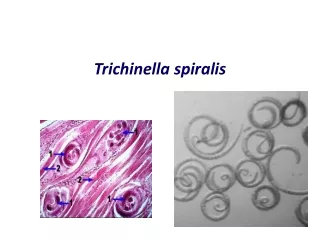
DIAGNOSIS Blood eosinophilia develops in >90% of patients with symptomatic trichinellosis and may peak at a level of >50% 2–4 weeks after infection. Serum levels of muscle enzymes, including creatinephosphokinase, are elevated in most symptomatic patients. Patients should be questioned thoroughly about their consumption of pork or wild animal meat and about illness in other individuals who ate the same meat. A presumptive clinical diagnosis can be based on fevers, eosinophilia, periorbital edema, and myalgias after a suspect meal. A rise in the titer of parasite-specific antibody, which usually does not occur until after the third week of infection, confirms the diagnosis. Alternatively, a definitive diagnosis requires surgical biopsy of at least 1 g of involved muscle; the yields are highest near tendon insertions. The fresh muscle tissue should be compressed between glass slides and examined microscopically (Picture 2), because larvae may be missed by examination of routine histopathologic sections alone. (Harrison’s Principles of Internal Medicine , 17 Edition Volume 1, p.1317) (Harrison’s Principles of Internal Medicine , 18 Edition)
TREATMENT Most lightly infected patients recover uneventfully with bed rest, antipyretics, and analgesics. Glucocorticoids like prednisone (Picture 3) are beneficial for severe myositis and myocarditis. Mebendazole and albendazole are active against enteric stages of the parasite, but their efficacy against encysted larvae has not been conclusively demonstrated. (Harrison’s Principles of Internal Medicine , 17 Edition Volume 1, p.1317) (Harrison’s Principles of Internal Medicine , 18 Edition)
PREVENTION Larvae may be killed by cooking pork until it is no longer pink or by freezing it at –15°C for 3 weeks. However, Arctic T. nativa larvae in walrus or bear meat are relatively resistant and may remain viable despite freezing. (Harrison’s Principles of Internal Medicine , 17 Edition Volume 1, p.1317) (Harrison’s Principles of Internal Medicine , 18 Edition)