Download
1 / 34
340 likes | 722 Views
Biology 130 – Molecular Biology and Genetics. Chromosomes dividing during Cell division. Kandinsky – Several Circles. Biology 130 – Molecular Biology and Genetics Knox College Winter 2007 Instructors: Matt Jones-Rhoades Stuart Allison

E N D
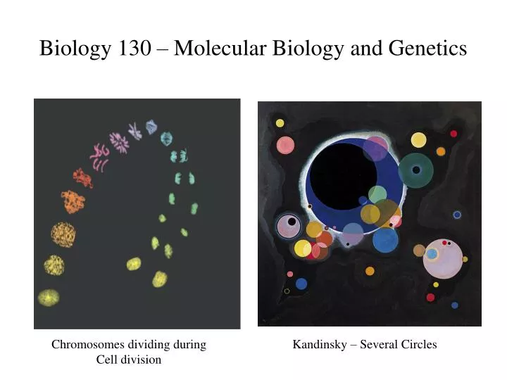
Biology 130 – Molecular Biology and Genetics Chromosomes dividing during Cell division Kandinsky – Several Circles
Biology 130 – Molecular Biology and Genetics Knox College Winter 2007 Instructors: Matt Jones-Rhoades Stuart Allison SMC B110 SMC B210 x7477 x7185 email: mjrhoade email: sallison Lab Coordinator: Ramiya Venigalla, SMC B113, x7386, email: rvenigal Office Hours: Jones-Rhoades: Tues 2nd Period, Wed 4th Period, Fri 6th Period. Allison: MWThF 3rd Period; Venigalla – MW 12:30-1:30 Lecture: SMAC A110 MWF 2nd Period Lab: SMAC B121 Textbooks: Campbell and Reece. 2011. Biology 9th Ed. Benjamin Cummings. Ridley. 2006. Genome. Perennial. Course Webpage: http://courses.knox.edu/bio130
Cell Division • Unicellular organisms • Reproduce by cell division • Multicellular organisms depend on cell division for • Development from a fertilized cell • Growth • Repair • The cell division process • Is an integral part of the cell cycle
Cell theory of life • ‘Where a cell exists, there must have been a preexisting cell, just as the animal arises only from an animal and the plant only from a plant.’ - Rudolf Virchow, 1855
Cell division results in genetically identical daughter cells • Cells duplicate their genetic material before they divide, ensuring that each daughter cell receives an exact copy of the genetic material, DNA • A cell’s endowment of DNA, its genetic information, is called its genome • The DNA molecules in a cell are packaged into chromosomes
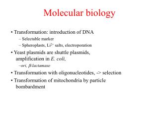
Chromosomes • Eukaryotic chromosomes • Consist of chromatin, a complex of DNA and protein that condenses during cell division • In animals • Somatic cells have two sets of chromosomes • Gametes have one set of chromosomes • In preparation for cell division • DNA is replicated and the chromosomes condense
Cell Division • Eukaryotic cell division consists of • Mitosis, the division of the nucleus • Cytokinesis, the division of the cytoplasm • In meiosis • Sex cells are produced after a reduction in chromosome number
The cell cycle consists of the mitotic phase and interphase. Interphase can be broken down into three phases – G1, S, and G2.
The Cell Cycle • We typically divide interphase into three phases – the G1 phase (for Gap 1), the S phase (for synthesis), and G2 phase (for gap 2). • The cell only duplicates its chromosomes (DNA) during the S synthesis phase. Thus a cell grows (G1), continues to grow as it synthesizes DNA and duplicates chromosomes (S), grows more and completes preparations for cell division (G2) and then divides (M). • Daughter cells then repeat the cycle – potentially infinitely.
By late interphase, the chromosomes have been duplicated but are loosely packed. • The centrosomes have been duplicated and begin to organize microtubules into an aster (“star”). Fig. 12.5a
In prophase, the chromosomes are tightly coiled, with sister chromatids joined together. • The nucleoli disappear. • The mitotic spindle begins to form and appears to push the centrosomes away from each other toward opposite ends (poles) of the cell. Fig. 12.5b
During prometaphase, the nuclear envelope fragments and microtubules from the spindle interact with the chromosomes. • Microtubules from one pole attach to one of two kinetochores, special regions of the centromere, while microtubules from the other pole attach to the other kinetochore. Fig. 12.5c
The spindle fibers push the sister chromatids until they are all arranged at the metaphase plate, an imaginary plane equidistant between the poles, defining metaphase. Fig. 12.5d
At anaphase, the centromeres divide, separating the sister chromatids. • Each is now pulled toward the pole to which it is attached by spindle fibers. • By the end, the two poles have equivalent collections of chromosomes. Fig. 12.5e
At telophase, the cell continues to elongate as free spindle fibers from each centrosome push off each other. • Two nuclei begin to form, surrounded by the fragments of the parent’s nuclear envelope. • Chromatin becomes less tightly coiled. • Cytokinesis, division of the cytoplasm, begins. Fig. 12.5f
Movement of chromosomes – In this model a chromosome tracks along a microtubule as the microtubule depolymerizes at its kinetochore end, releasing tubulin subunits – Pac-man mechanism.
Nonkinetichore (polar) microtubules are responsible for lengthening the cell along the axis defined by the poles. • These microtubules interdigitate across the metaphase plate. • During anaphase motor proteins push microtubules from opposite sides away from each other. • At the same time, the addition of new tubulin monomers extends their length.
Dinoflagellate Diatoms
A molecular control system drives the cell cycle • The cell cycle appears to be driven by specific chemical signals in the cytoplasm. • Fusion of an S phase cell and a G1 phase cell induces the G1 nucleus to start S phase. • Fusion of a cell in mitosis with one in interphase induces the second cell to enter mitosis. Fig. 12.12
The distinct events of the cell cycle are directed by a cell cycle control system. • These molecules trigger and coordinate key events in the cell cycle. • The control cycle has a built-in clock, but it is also regulated by external adjustments and internal controls. Fig. 12.13
A checkpoint in the cell cycle is a critical control point where stop and go signals regulate the cycle. • Many signals registered at checkpoints come from cellular surveillance mechanisms. • These indicate whether key cellular processes have been completed correctly. • Checkpoints also register signals from outside the cell. • Three major checkpoints are found in the G1, G2, and M phases.
For many cells, the G1 checkpoint, the restriction point in mammalian cells, is the most important. • If the cell receives a go-ahead signal, it usually completes the cell cycle and divides. • If it does not receive a go-ahead signal, the cell exits the cycle and switches to a nondividing state, the G0 phase. • Most human cells are in this phase. • Liver cells can be “called back” to the cell cycle by external cues (growth factors), but highly specialized nerve and muscle cells never divide.
Rhythmic fluctuations in the abundance and activity of control molecules pace the cell cycle. • Some molecules are protein kinases that activate or deactivate other proteins by phosphorylating them. • The levels of these kinases are present in constant amounts, but these kinases require a second protein, a cyclin, to become activated. • Levels of cyclin proteins fluctuate cyclically. • The complex of kinases and cyclin forms cyclin-dependent kinases (Cdks).
Cyclin levels rise sharply throughout interphase, then fall abruptly during mitosis. • Peaks in the activity of one cyclin-Cdk complex, MPF, correspond to peaks in cyclin concentration. Fig. 12.14a
MPF (“maturation-promoting factor” or “M-phase-promoting-factor”) triggers the cell’s passage past the G2 checkpoint to the M phase. • MPF promotes mitosis by phosphorylating a variety of other protein kinases. • MPF stimulates fragmentation of the nuclear envelope. • It also triggers the breakdown of cyclin, dropping cyclin and MPF levels during mitosis and inactivating MPF. Fig. 12.14b
The key G1 checkpoint is regulated by at least three Cdk proteins and several cyclins. • Similar mechanisms are also involved in driving the cell cycle past the M phase checkpoint.