
Introduction
Optically detected electron spin-flip resonance in CdMnTe S. Zeng, L. C. Smith, J. J. Davies, D. Wolverson, S. J. Bingham and G. N. Aliev Department of Physics, University of Bath, Bath, BA2 7AY, UK .

Introduction
E N D
Presentation Transcript
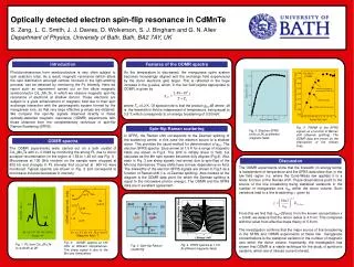
Optically detected electron spin-flip resonance in CdMnTeS. Zeng,L. C. Smith, J. J. Davies,D. Wolverson, S. J. Bingham and G. N. Aliev Department of Physics, University of Bath, Bath, BA2 7AY, UK Photoluminescence from semiconductors is very often subject to spin selection rules. As a result, magnetic resonance (which alters the spin distribution amongst centres involved in the light-emitting process) can be detected by monitoring the PL intensity. Here we report such an experiment carried out on the dilute magnetic semiconductor Cd1-xMnxTe, in which we observe magnetic spin-flip resonance of electrons at shallow donors. These electrons are subject to a giant enhancement of magnetic field due to their spin exchange interaction with the paramagnetic system formed by the manganese ions, so that very large effective g-values are obtained. We compare the spin-flip signals observed directly in these optically-detected magnetic resonance (ODMR) experiments with those obtained from the complementary technique of spin-flip Raman Scattering (SFRS). As the temperature is decreased, the manganese spins system becomes increasingly aligned and the exchange field experienced by the donor electrons gets larger. This is reflected in the huge increase in the g-value, which, in the low field regime appropriate to ODMR, is given by where T0=0.2 K. Of special note is that the product geff.DBwhere DBis the linewidth in field is independent of temperature, being equal to 3.5 T, which corresponds to an energy broadening of 0.20meV. Introduction Features of the ODMR spectra In SFRS, the Raman shift corresponds to the Zeeman splitting of the scattering centre, in this case the electron bound to a shallow donor. This provides the usual method for determination of gef.f. The electron SFRS spectra (blue arrow) at 1.5 K for a range of magnetic fields are shown in Fig.3. The shift is initially linear in field, but saturates as the Mn spin system becomes fully aligned (Fig.4). Also seen in Fig. 3 are sharp signals (red arrow) due to spin-flips of the Mn ions themselves. These shifts have a linear dependence on field. The linewidths of the electron SFRS signals are shown in Fig.5 as a function of Raman shift (i.e. of Zeeman splitting). Also marked on the diagram is the ODMR data point (for which the Zeeman splitting is equal to the microwave photon energy). The ODMR and the SFRS data are in excellent agreement. Fig. 5. FWHM of the SFRS signals as a function of Raman shift (Zeeman splitting). The ODMR data are shown by the intersection of the broken lines. Spin-flip Raman scattering Fig. 5. Electron SFRS shifts at 2K at different magnetic fields The ODMR experiments were carried out on a bulk crystal of Cd1-xMnxTe with x= 0.0048 by monitoring the strong PL due to donor acceptor recombination (in the region of 1.58 to 1.48 eV, see Fig. 1) . Microwaves at 105 GHz incident on the sample were chopped at 605 Hz and changes in PL intensity that occurred at 605 Hz were monitored. Typical spectra are shown in Fig. 2 and correspond to microwave-induced decreases in intensity. ODMR spectra The ODMR experiments show that the linewidth (in energy terms) is independent of temperature and the SFRS data show that, in the low field region (i.e. where the Curie-Weiss law applies) it is a linear function of the Raman shift. These observations point to the source of the line broadening being statistical variations in the number of manganese ions nMn within the donor volume. Such variations lead to a line broadening g given by From this we find that nMn=29 and, from the known concentration x = 0.048, we deduce that the donor radius is 4.5 nm. This compares with the value from effective mass theory of 5.8 nm. The investigation confirms that the major source of line broadening in the SFRS and ODMR experiments at these low manganese concentrations is the statistical variation in the number of magnetic ions within the donor volume. Importantly, the investigation has shown that ODMR is a viable technique for the study of spintronic systems, which are of intense current interest. Discussion Fig. 2. ODMR spectra at 105 GHz at different temperatures. The sharp signal is due to the Mn ions themselves. Fig. 1. PL from Cd1-xMnxTe (x=0.0045) at 2K Fig. 4. SFRS spectra at 1.5 K At different magnetic fields Fig. 3. Spin-flip Raman scattering


