Download
1 / 18
180 likes | 405 Views
Typical Spinal Nerve. Sanjaya Adikari Department of Anatomy. Skull. CNS is covered by the skull and the vertebral column PNS is outside. Vertebral column. Posterior median septum. Posterior white column. Posterior horn. Posterior nerve root. Lateral white column. Lateral horn.

E N D
Typical Spinal Nerve Sanjaya Adikari Department of Anatomy
Skull • CNS is covered by the skull and the vertebral column • PNS is outside Vertebral column
Posterior median septum Posterior white column Posterior horn Posterior nerve root Lateral white column Lateral horn Central canal Anterior nerve root Anterior horn Anterior white column Anterior median fissure Structure of a spinal cord segment
Dorsal root ganglion Somatic Autonomic
Dorsal and ventral roots • Dorsal roots contain afferent (sensory) axons • Ventral roots contain efferent (motor) axons • The ventral roots continue out from the spinal cord, and mix with their corresponding dorsal nerve root at a point after the ganglion • The combined dorsal and ventral roots are called a spinal nerve (therefore, spinal nerves are mixed).
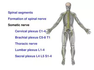
Spinal nerve is mixed (motor + sensory + autonomic) • Spinal nerve refers to the mixed spinal nerve • It is formed from the dorsal and ventral roots • Passes out through the intervertebral foramen • There are 31 bilaterally-paired spinal nerves • 8 cervical nerves (C1-C8) • 12 thoracic nerves (T1-T12) • 5 lumbar nerves (L1-L5) • 5 sacral nerves (S1-S5) • 1 coccygeal nerve (Co)
8 7 12 12 5 5 5 5 1 Spinal cord Vertebral column
C1 C1 C1 C2 C3 C4 C5 C6 C7 C8 C7 C8 T3 T1 T1 T2 T2 T3 T4 T5 T6 T10 T7 T8 T9 L1, L2 T10 T10 L3, L4 T11 L5 T12 S, C L1 Spinal nerves
Anterior and posterior primary rami • The posterior primary rami have lateral and medial branches They supply • back muscles and skin over the back • The anterior primary rami give off anterior and lateral cutaneous branches They supply • the rest of the body wall • Anterior primary rami also give rise to the roots of the various nervous plexuses
Posterior Lateral cutaneous branch Anterior cutaneous branch Anterior Medial Lateral Spinal nerves in the thoracic region These are typical spinal nerves
Rami communicantes • Gray ramus communicans • unmyelinated • White ramus communicans • myelinated
Nerve plexus Formedby communicating branches between anterior primary rami ofspinal nerves. Anterior primary rami form the roots of nerve plexus What are the advantages of a nerve plexus? Last’s Anatomy, 10th ed. Page 13
Dermatome Area of skin supplied by a single spinal nerve or spinal cord segment Myotome The muscle/s supplied by a single spinal nerve or spinal cord segment