
The Ear
E N D
Presentation Transcript
In the name of God The Ear Dr. Zahiri
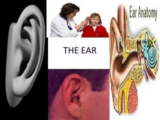
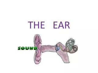
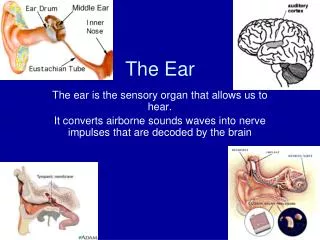
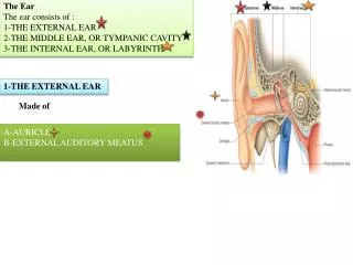
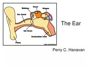
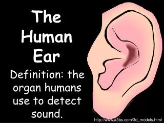
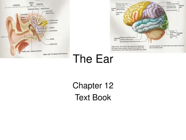
The Ear • is responsible of hearing and balance • This organ is composed of three regions, external ear, middle ear, and inner ear • External ear received sound and translated sound into mechanical vibrations • Middle ear amplified mechanical vibrations • Inner ear regulate hearing and maintain equilibrium • Vestibulocochlear nerve transmits sensory signals to the brain Dr. Maria Zahiri
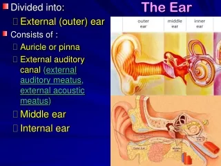
External Ear • The external ear is composed of : • the auricle or pinna • external auditory meatus • tympanic membrane Dr. Maria Zahiri
The auricle is composed of a plate of elastic cartilage covered by thin skin • The initial segment of external auditory meatus is composed of elastic cartilage that is continuous with the cartilage of pinna • Inner 2/3 of meatus is composed of bone • External auditory meatus • is covered with thin skin • containing hair, sebaceous • glands and modified sweat • glands known as ceruminous • glands that secrete earwax Dr. Maria Zahiri
Cerumen • A mixture of relatively viscous secretions from the sebaceous glands and non-viscous secretion from the ceruminous glands (modified apocrine glands) Dr. Maria Zahiri
Tympanic membrane covers the deepest end of the external auditory meatus • External surface of tympanic membrane is covered by a thin epidermis • internal surface is lined by a simple squamous or low cuboidal epithelium • The core of membrane is composed of a thin layer of connective tissue elements Dr. Maria Zahiri
Middle Ear • The tympanic cavity which is an air filled space in temporal bone is covered by simple squamous or low cuboidal epithelium • Three auditory ossicles located here are composed of compact bone • The ossicles covered by simple squamous epithelium and articulate together by synovial joints • Two skeletal muscles control motion between bone • Auditory (Eustachian) tube is lined by pseudostratified columnar ciliated epithelium Dr. Maria Zahiri
Eustachian (Auditory) Tube • Wall is bone closest to tympanic cavity, replaced by elastic cartilage except in nasopharynx where it becomes hyaline cartilage • Lined with ciliated epithelium, goblet cells and diffuse lymphoid tissue. • Equalizes air pressure between the tympanic cavity and the external environment. Walls normally closely apposed but open during swallowing and yawning. Dr. Maria Zahiri
Inner Ear • Inner ear is composed of bony labyrinth and membranous labyrinth • Bony Labyrinth • is part of temporal bone that is lined by endosteum • Perilymphatic space separated bony labyrinth from membranous labyrinth which is filled by a clear fluid known as perilymph • Membranous labyrinth suspended in this fluid • Central region of bony labyrinth is called vestibule that is between posteriorly placed semicircular canals and anteriorly placed cochlea Dr. Maria Zahiri
INNER EAR – A membranous labyrinth filled with endolymph inside a bony labyrinth filled with perilymph Dr. Maria Zahiri
Bony Labyrinth Dr. Maria Zahiri
Components of the Bony Labyrinth Vestibule: Contains Utricle and Saccule Semicircular Canals: Extend from vestibule. Superior, lateral and posterior at right angles to one another. Contains the semicircular ducts. At one end of each is a dilatation, the ampulla. Cochlea: Coils like a snail around a central pillar of bone, the modiolus. The cochlear duct rests upon a partial shelf of bone projecting from the modiolus, the osseous spiral lamina Dr. Maria Zahiri
Helicotrema Modiolus Osseous Spiral Lamina Dr. Maria Zahiri
Membranous Labyrinth Dr. Maria Zahiri
Sensory Regions Dr. Maria Zahiri
Saccule and Utricle • Saccule and Utricle are composed of a thin vascular CT that its inner side is lined by a simple squamous to low cuboidal epithelium consists of light and dark cells • Light cells may play a role in absorption of endolymph and dark cells control its composition • Macula is a thickened specialized region of epithelium which acts as receptor for linear acceleration • Maculae are composed of neuroepithelial cells (type I) and hair cells (type II) and supporting cells • Type I and II cells have a single kinocillium and about 100 stereocilia
Saccule and Utricle • Stereocilia arranged in rows according to length, the longest being nearest to kinocilium • Supporting cells maintain the hair cells and contribute to production of endolymph • Stereocilia of neuroepithelial cells are embeded in a thick gelatinous, glycoprotein membrane known as otolithic membrane • Surface of this membrane is contains calcium carbonate crystals known as otolithsor otoconia • Type I and II cells innervated by vestibular branch of VIII cranial nerve Type I: Large cup-shaped ending Type II: Many afferent endings
Saccule & Utricle Dr. Maria Zahiri
Semicircular ducts • These are located in semicircular canal, arising from utricle • Near the utricle ducts expanded and form ampullae which are contain cristae ampullares • Crista ampullaris is composed of a ridge that covered by sensory epithelium consists of supporting cells and neuroepithelial hair cells ( type I and II ) • Hair cells do not lie on basal lamina and are located between supporting cells • Cupula is similar to otolithic membrane but it is cone shape and has not otolith
Cochlear Duct • Cochlear duct contains organ of Corti that is responsible to responds to sound • Modiolus has a lateral projection which is known as osseous spiral lamina • Periosteum of the osseous spiral lamina forms spiral limbus • Spiral organ of Corti supported by the thickened periosteum of the cochlea that is called spiral ligament • Basilar membrane is a thin membrane that extends between the spiral ligament and the osseous spiral lamina
Basilar membrane support the organ of Corti and its vibration induced by disturbances in the perilymph detected by hair cells of the organ of Corti • Vestibular membrane extends across the cochlear duct • Vestibular Membrane is composed of two layers of flattened squamous epithelial cells • Stria vascularis is a pseudostratified epithelium that contains intraepithelial plexus of capillaries • Stria vascularis extends between vestibular membrane and the spiral prominence • Spiral prominence is a protuberance covered by epithelium that continuous with stria vascularis and continues onto basilar membrane that is called cells of Claudius
COCHLEA DUCT Scala Media is the space within the triangular shaped cochlear duct. It is filled with endolymph The cochlear duct and the spiral lamina divide the cochlea into two additional spaces: The scalavestibuliand the scala tympani. Both are filled with perilymphand are contiuous with one another at the helicotrema Dr. Maria Zahiri
Cells of Organ of Corti • It is composed of inner and outer hair cells and supporting cells • Supporting cells consist of: inner and outer pillar cells, inner and outer phalangeal cells and cells of Hensen • Inner hair cells are neuroepithelial and responsible in the reception of sound • They are arranged in a single row along the entire length of cochlea • Their stiff stereocilia arranged in a W-shaped formation but no kinocilium • At the base of each hair cell are efferent and afferent nerve endings that transmit impulses to the bipolar neurons of the spiral ganglion.
0 Cochlear Duct Dr. Maria Zahiri
Cells of Organ of Corti • Outer hair cells are also neuroepithelial are three or more rows located between outer phalangeal and outer pillar cells • Inner and outer pillar cells rest on basilar membrane, enclose the inner tunnel of Corti • Inner and outer phalangeal cells are intimately associated with the inner and outer hair cells respectively • They support nerve fibers that synapses with the hair cells
Tectorial Membrane • Tectorial membrane overlies the tip of hair cells • It is composed of glycoproteins
Endolypmphatic Duct and Sac • Simple squamous epithelial lining becoming columnar near the sac. • Columnar epithelium has two cell types, one with microvilli, pinocytotic vesicles and vacuoles. • This cells may be responsible for the absorption of endolymph and endocytosis of foreign materials Dr. Maria Zahiri
Path of sound waves – in through oval window – up scala vestibuli – through Helicotrema – along tympani and dissipation through round window 0 Dr. Maria Zahiri
Dr. Maria Zahiri با آرزوی بهروزی