Download

1 / 19
190 likes | 345 Views
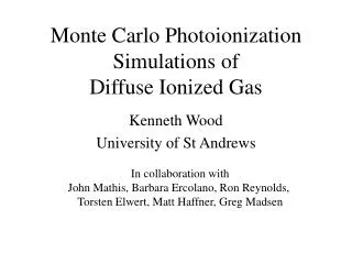
Multiple Photoionization of C 60. K. A. Barger, R. Wehlitz, and P. Juranic. Synchrotron Radiation. Electro Magnetic Radiation emitted by charged particles that are that are traveling at relativistic speeds and that are accelerated by magnetic fields

E N D
Multiple Photoionization of C60 K. A. Barger, R. Wehlitz, and P. Juranic
Synchrotron Radiation • Electro Magnetic Radiation emitted by charged particles that are that are traveling at relativistic speeds and that are accelerated by magnetic fields • The source of this radiation was the Aladdin electron storage ring at the Synchrotron Radiation Center (SRC) in Stoughton, Wisconsin.
Schematic of the Aladdin ring Port 042 6m TGM Undulators Bending Magnets
Flux vs. the Aladdin ring photon energy for SRC's bending magnets and undulators
Photoionization This is when a photon interacts with a particle causing it to lose one or more electrons and become positively charged e Photo-effect: Usually thought of as one photon being absorbed by the atom/molecule and one electron is emitted
Simultaneous emission • One photon comes in and causes two electrons to be simultaneously ejected through electron correlation • Coulomb Dipole interactions occur between the: • Emitted electrons • Remaining electrons • Nucleus of the atom e - + e e e
The Cross Section σ The ionization cross section is a measure of the probability that the particle will become ionized. Example of Rutherford Scattering Cross Sections
History of Double Photoionization In 1988, the first near threshold experiment was done on He. Wannier Theory: α=1.056 Experimental: α=1.05 ± 0.02 79 Other experiments included oxygen & sodium, but had: Large error bars Few photon energies
Recent Years Ralf Wehlitz has studied Li and Be He measured the double-to-single photoionization ratio with high accuracy near the threshold energy and has found oscillations in the double photoionization cross section Be Be2+’s relative cross section as a function of excess energy Excess Energy = Photon Energy – Threshold Energy
Double-Photoionization Cross Section of Beryllium Δσ is the Difference between our DPI cross section data and smooth theoretical Wannier curve Coulomb Dipole Theory
Photoionization of C60Experimental Setup TAC-measures the time difference between the PP and the time for the C60 ions to reach the MCP PP - PusherPlate EP - ExtractorPlate CP - Condenser plate MCP - Microchannel Plate CFD - Constant Fraction Discriminator TAC - Time to Amplitude Converter MCB – Multichannel Buffer MCB-sorts the pulse heights into channels which creates a spectrum CFD-used to cut off noise and it also gives pulse positions that are independent of the height of the pulses PP-Pushes all ions through the extractor plate by creating a localized electric field. The pulse applied to the pusher plate serves as the stare pulse of the Time-to-Flight measurement EP-a grounded plate marking the boundary of the localized electric field CP-improves the vacuum by freezing unwanted gases and un-ionized C60 to the surface of the plate MCP-an array of three detector plates that have voltages between 2800-3000 Volts. These Plates are designed to convert ionized particles into electric pulses, which can be used to count C60 ions
Time-of-Flight Mass Spectrometer Measures mass-to-charge ratio (m/q) which forms separate peaks for each charge state This can be used to find the Relative Ionization Cross-Section (atomic mass units/charge) This spectrum was taken using photons at an energy of 154eV and with the oven set to a temperature of 324°C.
Ratio of Ionization Charge States as a Function of Excess Energy Work done by Ralf Wehlitz in March of 2004
Oscillations in the C602+/ C60+ Cross-Section ratio Δσ is the Difference between our DPI cross section data and smooth theoretical Wannier curve Work done by Ralf Wehlitz in March of 2004
Ratio of Ionization Charge States as a Function of Excess Energy New The ratio of the integrated peak areas C603+/ C601+versus the excess energies The ratio of the integrated peak areas C602+/C601+ versus the excess energies
The Wannier Theory & Coulomb Dipole Theory: Only apply to near threshold They do not apply to molecules Strangely Coulomb Dipole Theory does correctly predict the oscillations in the cross sections for C60, but the theory applies to atoms Problems with Theories
Summary • Using a Time-of-Flight mass spectrometer we are able to studying the 1+ to 3+ charge states as a function of excess energy • This information can be used to determine the relative cross sections of each charge state • We have observed that the double ionization cross section ratio does not change linearly, and that the amplitude and wave length of the oscillations change with excess energy • The theories available only apply to atoms and not molecules
Acknowledgments I would like to thank the REU program at University of Wisconsin-Madison, and the staff of the Synchrotron Radiation Center for their support. I would also like to thank my mentor at the SRC Ralf Wehlitz, and Pavle Juranic as well as my advisor Jim Stewart at WWU for all their help and guidance. This work is based upon research conducted at the Synchrotron Radiation Center, University of Wisconsin-Madison, which is supported by the NSF under Award No. DMR-0084402
References: [1] D. Lukić, J. B. Bluett, and R. Wehlitz, Phys. Rev. Lett. 93, 023003 (2004). [2] R. Wehlitz, J. B. Bluett, and S. B. Whitfield, Phys. Rev. Lett. 89, 093002 (2002). [3] A. Reinköster, S. Korica, G. Prümper, J. Viefhaus, K. Godehusen, O. Schwarzkopf, M Mast, and U. Becker, Rev. Phys. B 37, 2135-2144 (2004). [4] H. Steger, J. de Vries, B. Kamke, W. Kamke, and T. Drewello, Chem. Phys. Lett. 194, 452-456 (1992). [5] R. K. Yoo, B. Ruscic, and J. Berkowitz, J. Chem. Phys. 96, 911-918 (1992). [6] R. Wehlitz, D. Lukić, C. Koncz, and I. A. Sellin, Rev. Sci. Instrum. 73, 1671-1673 (2002). [7] J. B. Bluett, D. Lukić, and R. Wehlitz, Phys. Rev. A 69, 042717 (2004). [8] J. M. Rost, Priv. Comm. (2004). [9] M. J. Seaton, J. Rev. Phys. B 20, 6363-6378 (1987). [10] S. Petrie, and D. K. Bohme, Rev. ApJ 540, 869-885 (2000). [11] SRC http://www.src.wisc.edu/ (2004). [12] J. J. Brehm, and W. J. Mullin, Introduction to the Structure of Matter (1989). [13] C. R. Nave, Rutherford scattering hyperphysics.phy-astr.gsu.edu/hbase/rutsca (2003).