Download
1 / 26
260 likes | 416 Views
Front Back. This tutorial will take you through the basic structure and function of the front part of the eye. It contains both information and questions to test yourself.

E N D
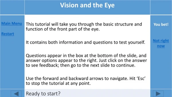
Front Back • This tutorial will take you through the basic structure and function of the front part of the eye. • It contains both information and questions to test yourself. • Questions appear in the box at the bottom of the slide, and answer options appear to the right. Just click on the answer to see feedback; then go to the next slide to continue. • Use the forward and backward arrows to navigate. Hit ‘Esc’ to stop the tutorial at any point. You bet! Not right now Ready to start?
Posterior chamber Does that word really refer to the front ? Check again! The pupil‘s in the middle of the iris. Good work! You figured it out. Pupil Anterior chamber Image modified from Wikipedia, https://en.wikipedia.org/wiki/Mammalian_eye used under a Creative Commons license The part of your eye in front of the iris is the _____
The sclera is the white of your eye. That’s right! The cornea’s completely transparent. The iris is inside your eye! Sclera Cornea Iris Image modified from Wikipedia, https://en.wikipedia.org/wiki/Mammalian_eye used under a Creative Commons license The clear layer covering the anterior chamber is the:
Sclera Light can’t pass through the sclera! The iris isn’t transparent – it’s the colored part! Good work! The lens lets light pass through. Iris Lens Image modified from Wikipedia, https://en.wikipedia.org/wiki/Mammalian_eye used under a Creative Commons license Light rays bend as they pass through the cornea and:
Sclera But no light can pass through the sclera! The cornea’s transparent! It can’t stop light. Right! The iris can adjust the pupil’s size to let light in. Cornea Iris Image modified from Wikipedia, https://en.wikipedia.org/wiki/Mammalian_eye used under a Creative Commons license The ____ controls how much light reaches the lens
Good work on the anatomy! • The anterior chamber of your eye is all about FOCUSING light. • Focusing means bringing the light rays that have bounced off one spot on an object together again, so you see them as a sharp spot instead of a fuzzy blur. Image modified from Wikipedia, https://en.wikipedia.org/wiki/Bokeh used under a Creative Commons license
When light hits an object, the light rays bounce off that object in all different directions. • They hit your cornea at different places, and different angles. Image from Pixabay, https://pixabay.com/en/light-ray-blaze-gradient-734436/ used under a Creative Commons license
The surface of the cornea bends the light rays toward the pupil. • How much of the light passes through the pupil is controlled by the iris. Dilates That would let more light in Right! So you don’t get too much light! If that happened, you couldn’t see. Constricts Closes entirely Image modified from Wikipedia, https://en.wikipedia.org/wiki/Mammalian_eye used under a Creative Commons license In bright light, the pupil _________
Do you see that the iris has radial fibers coming out from the pupil and concentric fibers going around the pupil? • These allow the pupil to constrict and dilate. Radial Right! The radial fibers will pull the pupil open Think again. These fibers go round like a drawstring. They’d cancel each other out! concentric Con-centric radial Radial and con-centric Image from Wikipedia, https://en.wikipedia.org/wiki/Iris_%28anatomy%29 Used under a Creative Commons license When the _______ fibers contract, the pupil will dilate.
Contracting the radial fibers pulls the pupil open farther, dilating it. • Contracting circular fibers pulls the pupil closed, like a drawstring. Radial That’s right! These fibers pull the pupil open. The SNS dilates the pupils. Will this do it? They’d cancel each other out! concentric Con-centric radial Radial and con-centric Image from Wikipedia, https://en.wikipedia.org/wiki/Iris_%28anatomy%29 Used under a Creative Commons license The sympathetic system (SNS) makes the ____ fibers contract
The sympathetic system stimulates the radial fibers, causing pupil dilation – the better to see the emergency with! Darkness has the same effect. • The parasympathetic system does the opposite, stimulating the concentric fibers and closing the pupil. Bright light has the same effect. concentric radial Image from Wikipedia, https://en.wikipedia.org/wiki/Iris_%28anatomy%29 Used under a Creative Commons license
The cornea only begins focusing the light. • The light rays are bent further toward one another as they pass through the lens. • One advantage of the lens is that its shape can be adjusted, so the amount it bends the light can be adjusted too. Think about it. Does flat window glass bend light? Right! The less lens curvature, the less light bends. If the lens changes, the light bending will change! More Less Just the same Image modified from Tessa Johnson, 2007 https://www.flickr.com/photos/tessawatson/412727556 Used under a Creative Commons license If the lens becomes flatter, light rays will bend ___ as they pass through it
A convex, curved lens bends light rays toward each other more than a flat piece of glass or a concave lens. • The lens in your eye never becomes completely flat – but it can become flatter, and then the light rays don’t bend as far toward each other. Image modified from Tessa Johnson, 2007 https://www.flickr.com/photos/tessawatson/412727665/ Used under a Creative Commons license
Right! You’ll need to bend the light a lot to bring those rays together. If the lens is too flat, those light rays won’t come together to focus. • Why would you ever want to make your lens flatter or rounder? • It has to do with the light rays entering your eye. • Here are light rays coming from a very close object into your eye. Look how slanted the top and bottom rays are! More rounded More flattened Image modified from Wikipedia, https://en.wikipedia.org/wiki/Mammalian_eye used under a Creative Commons license To focus these rays, your lens will need to be _____
It would take a pretty round lens to bend those diverging light rays back together. • This is what happens when you look at something really close to your face. Your lens gets rounder, to focus on it. Image modified from Wikipedia, https://en.wikipedia.org/wiki/Mammalian_eye used under a Creative Commons license
Now let’s move the object away. The light rays still spread • out … • But the • ones that spread • out too much don’t even hit your eye! Image modified from Wikipedia, https://en.wikipedia.org/wiki/Mammalian_eye used under a Creative Commons license
Do these light rays need to bend so much? Right! These light rays are less slanted and don’t need to bend as much. • The light rays that hit your eye are not very slanted. More rounded More flattened Image modified from Wikipedia, https://en.wikipedia.org/wiki/Mammalian_eye used under a Creative Commons license To focus on an object far away, the lens should be:
See how much more the light from the close object must bend, to bring the rays back together?
If you pull on the edges, you will stretch the lens out. Right! You’ll pull the lens out flatter. • So: • When you look at something close, your lens needs to be rounded. • When you look at something far away, your lens needs to be flattened. • How do you change its shape? By pulling on the edge. More rounded More flattened Pulling on the edge of your lens would make it ____
You’ll be pulling it out flat! Right! You’ll be pulling it out flat. • Here’s one edge of your lens. See the cornea and iris in front of it? • Now, see the strands attached to the edge? • These are SUSPENSORY LIGAMENTS. One end attaches to the edge of your lens, and the other end attaches to a ring of muscle – the CILIARY MUSCLE. Get rounder Get flatter CILIARY MUSCLE SUSPENSORY LIGAMENTS Image modified from Wikipedia, https://en.wikipedia.org/wiki/Ciliary_processes used under a Creative Commons license When you pull on the suspensory ligaments, the lens will:
Good! If the ring of muscle is smaller, it won’t pull on the ligamentsas much. Recheck. If the ring is larger, won’t it pull on the ligaments? • Let’s look at it from the front. • Here’s the ring of ciliary muscle, with the suspensory ligaments holding the lens in position, and pulling on the edge of the lens. • The ciliary muscle is made of concentric fibers. When they contract, the ring of ciliary muscle gets smaller. When they relax, it gets larger. Contract Relax LENS So to make your lens rounder, you would have to ___ the ciliary muscle fibers.
Contracting the ciliary muscle fibers will make the ring of muscle smaller. • It won’t pull so hard on the edge of the lens – • And the lens will be able to bulge up and get rounder. LENS
To summarize: • Close objects need a rounder lens, so you must contract the ciliary muscle. • Far-away objects need a flatter lens, so you relax the ciliary muscle. • You know that feeling of eye-strain you get when you’re looking at something close-up? That’s your ciliary muscle getting tired. Then you give it a rest by looking at something far away!
We’ve gone over the functions of the cornea, iris, lens, and ciliary muscle. • But what about the stuff that fills the anterior chamber – the aqueous humor? • This watery fluid leaks out of blood vessels and carried food and O2 to the transparent structures of your eye, where blood vessels would block the light.
Aqueous humor is made by tiny vessels in the base of the ciliary muscle. • It flows over the surface of the lens, between the lens and the iris, and out into the front of the eye where it carries nutrients and O2 to the cornea. Then it’s reabsorbed into the ring-shaped Canal of Schlemmwhich lies in the base of the iris. CILIARY MUSCLE SUSPENSORY LIGAMENTS Image modified from Wikipedia, https://en.wikipedia.org/wiki/Ciliary_processes used under a Creative Commons license
Too much aqueous humor can cause high pressure in the eye, called glaucoma. • This is the end of the review of the front of the eye – the part that deals with focusing images and controlling the amount of light that enters the pupil. • Now you should be ready to look at the back of the eye – the part where nerve cells actually fire in response to that light, sending messages to your brain!