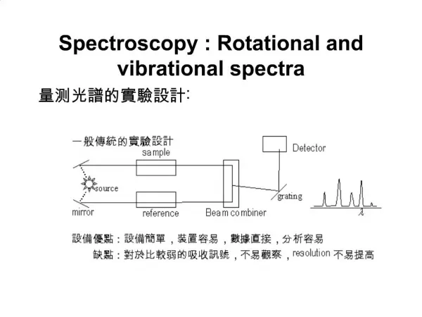
Vibrational Spectroscopy for Pharmaceutical Analysis
Vibrational Spectroscopy for Pharmaceutical Analysis . Part IV. Fourier Transform Infrared (FT-IR) Spectroscopy Rodolfo J. Romañach, Ph.D. LAB INSTRUMENTS. Spectrum One – Perkin Elmer. Scimitar-Varian . Thermo Nicolet 6700. Tensor 27 – Bruker Optics. ABB - 100. PROCESS INSTRUMENTS.

Vibrational Spectroscopy for Pharmaceutical Analysis
E N D
Presentation Transcript
Vibrational Spectroscopy for Pharmaceutical Analysis Part IV. Fourier Transform Infrared (FT-IR) Spectroscopy Rodolfo J. Romañach, Ph.D.
LAB INSTRUMENTS Spectrum One – Perkin Elmer Scimitar-Varian Thermo Nicolet 6700 Tensor 27 – Bruker Optics ABB - 100
PROCESS INSTRUMENTS ABB-200 Hamilton Sundstrand RefinIR Mettler Toledo ReactIR™4000 ABB-400
Instrumentation • The vast majority of modern infrared spectrometers are Fourier Transform Infrared Spectrometers.
Advantages of FT-IR Spectroscopy • Obtains the information on all the frequencies at the same time (Multiplex or Fellgett Advantage) and as a result: • Signal to noise ratio may be improved by increasing number of scans by N1/2. • Fast, may be used for dynamic processes: GC/FT-IR. Fellgett or Multiplex Advantage
Advantages of FT-IR Spectroscopy • Radiation may be moved away from optical bench to interface instrument to IR microscope or GC/FT-IR. • Provides precise and accurate determination of absorption wavelength. Frequency calibration with He-Ne laser.
Schematic of a Michelson Interferometer. Figure 8, page 87 Chalmers and Dent.
Schematic showing summation of two cosine function interferogram. Chalmers and Dent page 88.
An IR spectrum can be obtained, after applying the Fourier Transform.
Essential Components of FT-IR Spectrometer • Interferometer (KBr/Ge beamsplitter used for mid-IR, CaF2/Fe2O3 for NIR). • Computer. • He:Ne laser. • Source. • Mirrors. • Detectors.
Detectors • DTGS (Deuterium Trygliceride Sulfate) – most commonly used, can be used at room temperature without cooling. Detector by default, DTGS will be included in new FT-IR spectrometer, unless MCT is requested. • Mercury Cadmium Telluride (HgCdTe, or MCT) – have higher sensitivity and higher response speed than DTGS and are used in IR microscopes and in GC/FT-IR experiments. Liquid nitrogen cooling required. Non-linear at higher radiation intensities. H. Günzler, H. U. Gremlich, IR Spectroscopy An Introduction, Wiley-VCH, 2002, pages 60 – 61, 70
Fourier transform from space domain to frequency domain. B(ν) is the single beam spectrum, δ is the optical retardation.
For a polychromatic source, the interferogram represents the summation of all the individual cosine functions corresponding to each of the wavelengths (wavenumbers in the source). They are only in phase at the centerburst (position of zero path difference.
Sequence for Obtaining Spectrum • Interferogram of Background is obtained (without sample) • System uses Fourier Transform to create single beam background spectrum. • Interferogram of Sample is obtained. • System uses Fourier Transform to create single beam spectrum of sample. • System calculates the transmittance or absorbance spectrum.
The CO2 Band • If the CO2 concentration is the same for the background and sample spectra it will be ratioed out. • Should have a stream of dry air going through the sample compartment to keep CO2 constant. If air stream is too high, it could cause disturbance in sample compartment. • When sample compartment is open, CO2 comes in, wait a few seconds to re-establish purge and take spectrum.
Methyl Heptanoate Problem with CO2 Band.
100% line test • The efficiency of the spectrometer may be checked with successive single beam spectra of the empty sample compartment. • The spectrum should be a straight line, but some spectral noise will always be observed. • This test also serves to document the spectral noise. H. Günzler, H. U. Gremlich, IR Spectroscopy An Introduction, Wiley-VCH, 2002, pages 67-69
Strong CO2 and water bands but the baseline is flat, instrument is working well.
Wavelength Accuracy • The He:Ne laser monitors the position of the moving and triggers data acquisition. • The He:Ne laser serves as an internal reference for every interferogram obtained. • “The abscissa precision of a data point in a FT spectrum recorded on a commercial spectrometer is usually quoted as better than 0.01 cm-1” page 97 Chalmers and Dent. • You will see a yellow-red beam, this is the source and He:Ne laser together.
Accuracy in wavelength determination, because of He:Ne laser.
Source of Radiation • Laser is not the source of radiation. • Globar is most commonly used (silicon carbide rods). At a temperature of 1500 K, it provides substantial energy. • Other instruments use chromium nickel alloy wires, or nerst rod (zirconium oxide) that when heated emit radiation. • Sources may last several years, not necessary to turn them off. H. Günzler, H. U. Gremlich, IR Spectroscopy An Introduction, Wiley-VCH, 2002, page 40.
Choice of Parameters for Spectral Acquisition The following parameters must be taken into consideration when taking spectra: 1. Apodization & Number of Points • Resolution • Number of Spectra
The moving mirror does not travel to plus or minus infinity, so the interferograms is truncated (cut) by a boxcar function. Schematic of Boxcar Truncation of the interferogram for a single frequency source. Chalmers and Dent figure 11, page 90.
Software will have a command requesting how many points you want to keep away from centerburst. Most of the information is in first 100 points, but most users keep 300 – 500 points. System has ADC with dynamic range of 1016 or better.
The Fourier Transform is now a sinc function and positive and negative lobes are introduced to each band. Effect of Boxcar Truncation Effect of Boxcar Truncation You might choose double sided interferogram and 300 points, you are specifying the truncation of the spectrum.
Apodization • The lobes may be eliminated by replacing the boxcar function with another function. • However, this process always increases the spectral band width and decreases band intensity. • The functions are called apodization functions. • The IR spectrometer software will give you a wide range of choices.
There is a tradeoff between eliminating the lobes and having band widening with reduction in intensity. • However, apodization is used in most experiments. • The weak Norton-Beer function is probably the most commonly used apodization function. Effect of Triangular Apodization.
Spectral Resolution • The spectral resolution is related to the travel of the moving mirror. • To resolve two bands in the spectrum, the mirror must travel to complete one beat pattern generated between the two cosine waves in the interferogram that represent the wavenumber portions of the spectrum. • Most FT-IR spectrum can provide resolution of at least 2 cm-1. • For liquids usually 8 cm-1 resolution is sufficient, some solids may require 4 cm-1 resolution. • The spectral bandwidths could be much greater than the resolution.
Spectral Resolution • At higher resolutions (2 cm-1) you will see more noise in the spectrum than at a lower resolution (8 cm-1).
Number of Spectra • Use multiplex advantage to improve SNR (signal to noise ratio) of spectra. • Take at least 16 spectra for a sample to improve SNR by a factor of 4. • If you take 64 spectra at a spectral resolution of 8 cm-1, you can improve SNR by a factor of 8, and the total spectral collection time is about 1 minute.