Download
1 / 61
620 likes | 898 Views
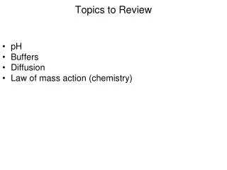
Topics to Review. pH Buffers Diffusion Law of mass action (chemistry). Functions of the Respiratory System. Provides a way to exchange O 2 and CO 2 between the atmosphere and the blood oxygen is used by the cells of the body solely for the process of aerobic respiration

E N D
Topics to Review pH Buffers Diffusion Law of mass action (chemistry)
Functions of the Respiratory System Provides a way to exchange O2 and CO2 between the atmosphere and the blood oxygen is used by the cells of the body solely for the process of aerobic respiration carbon dioxide is a waste product of aerobic respiration and must be removed from the body Regulation of body pH Protection from inhaled pathogens and irritating substances Vocalization
The Respiratory System Together, the respiratory system and the circulatory system deliver O2 to cells and remove CO2 from the body through 3 processes Pulmonary ventilation (breathing) movement of air into and out of the lungs Inspiration/inhalation and expiration/expiration Gas Exchange O2 and CO2 are exchanged between the air in the lungs and the blood O2 and CO2 are exchanged between the blood and the cells Transport movement of O2 and CO2between the lungs and cells
Organization of the Respiratory System • Anatomically, the respiratory system includes the: • upper respiratory tract (mouth, nasal cavity, pharynx and larynx) • lower respiratory tract (the trachea, 2 primary bronchi, the branches of the primary bronchi and the lungs) • Functionally, the respiratory system includes the: • the conducting zone (semi-rigid airways) lead from the external environment of the body to the exchange surface of the lungs • the exchange surface (respiratory zone) consists of the alveoli which are a series of interconnected sacs (surrounded by pulmonary capillaries) which expand and collapse during ventilation and allows oxygen and carbon dioxide to be exchanged between the air in the lungs and the blood
The Thorax and Respiratory Muscles The bones of the spine and ribs and their associated skeletal muscles form the thoracic cage Contraction and relaxation of these muscles alter the dimensions of the thoracic cage which promotes ventilation 2 sets of intercostal muscles connect the 12 pairs of ribs additional muscles (sternocleidomastoid and scalenes) connect the head and neck to the sternum and the first 2 ribs a dome-shaped sheet of skeletal muscle called the diaphragm forms the floor the abdominal muscles also participate in ventilation
The Pleural Membranes and Fluid Within the thorax are 2 double layered pleural sacs surrounding each of the 2 lungs Parietal pleura lines the interior of the thoracic wall and the superior face of the diaphragm Visceral pleura covers the external surface of the lungs (alveoli) A narrow intrapleural space between the pleura is filled with 25 mL of pleural fluid which holds the 2 layers together by the cohesive property of water serves to lubricate the area between the thorax and the outer lung surface holds the lungs tight against the thoracic wall prevents lungs from completely emptying even after a forceful expiration
Proximal Respiratory Tract Air enters the upper respiratory tract through either the mouth or nose and passes through the pharynx warms and humidifies (adds H2O) inspired air hair in the nose filters inspired air of any dust Air then passes through the larynx or“voice box” contains the vocal cords (bands of connective tissue) which tighten and vibrate to produce sound Air continues into the lower respiratory tract through the trachea which is a semi-flexible tube held open by C-shaped rings of cartilage The distal end of the trachea splits into 2 primary bronchi which lead to the 2 lungs branch repeatedly into progressively smaller bronchi the walls of the bronchi are supported by cartilage
The inner (mucosal) surface of the trachea and bronchi consists of epithelial tissue that functions as the mucocilliary escalator to trap and eliminate debris Goblet cells secrete mucus to trap debris in inspired air Pseudostratified ciliated columnar epithelium move debris trapped in mucus up towards the mouth for expectoration/swallowing
Middle and Distal Respiratory Tract Bronchi send air into the bronchioles these airways are supported by smooth muscle only contraction causes bronchoconstriction which decreases the airway diameter and makes ventilation more difficult increases airway resistance to decrease flow relaxation causes bronchodilationincreases the airway diameter which makes ventilation easier decreases airway resistance to increase flow branch into respiratory bronchioles which begins the Bronchioles move air into the blind sacs called alveoli where gas exchange occurs (respiratory zone) approximately 150 – 300 million per lung
Anatomy of Alveoli Composed of very thin(simple)epithelial tissue consisting of 2 predominant alveolar cell types Type I (squamous)alveolar cells allows for very rapid exchange of O2 and CO2 Type II or great (cuboidal) alveolar cells secrete surfactant into the alveolar lumen Exterior surface is surrounded by large numbers of blood capillaries for gas exchange and large numbers of elastic fibers to aid in lung recoil during exhalation White blood cells (macrophages) within the lumen of the alveoli protect against inhaled pathogens Alveoli represents an enormous surface area for gas exchange (2800 square feet or half of a football field)
Properties of Alveoli Compliant ability to be easily stretched or deformed allows lungs to fill up with air during inspiration attributed by the very thin Type I alveolar cells Elastic ability to resist being stretched or deformed allows lungs recoil (deflate) during expiration attributed by: interior (luminal) surface covered with a thin film of water which creates surface tension at the air-fluid interface (surface) of the alveoli the elastic fibers surrounding the alveoli
Alveolar Surface Tension and Elasticity During inhalation the alveoli expand and adjacent water molecules on the luminal surface are pulled apart from one another causing the H-bonds between them to be stretched (like a spring) creating tension During exhalation the tension within the H-bonds is released which returns the water molecules to their original spacing pulling the alveoli inward allowing them to recoil
Surfactant Type II alveolar cells secrete surfactant (“surface active agent”) which is a fluid consisting of amphiphilic molecules into the lumen of the alveoli These molecules disrupt the cohesive forces between water molecules by inserting themselves between some of the water molecules preventing H-bonds from forming and thus decreases the surface tension of the water on the luminal surface Reducing surface tension simultaneously increases compliance and reduces elasticity of the alveoli which greatly decreases the amount of effort needed to inflate the lungs while retaining the ability to deflate the lungs Without surfactant, the muscles of respiration cannot contract with enough force to overcome the alveolar surface tension resulting in the inability to breathe
Pulmonary Ventilation The movement of air into and out of the airways occurs as a result of increasing and decreasing the dimensions of the thoracic cavity through the contraction and relaxation of the skeletal muscles of respiration Since the alveoli are “stuck” to the interior surface of the thorax via the pleura, dimensional changes in the thoracic cavity result in the same dimensional changes in the alveoli Dimensional changes in the alveoli create air pressure changes in the alveoli as expressed by Boyle’s Law
Boyle’s Law The mathematical inverse relationship that describes what happens to the pressure of a gas or fluid in a container following a change in the volume (dimensions) of the container If the volume of a container increases, then pressurewithin the container mustdecrease If volume of a container decreases, then pressure within the container mustincrease V1xP1= V2xP2 V = volume of a container P = pressure within the container force of collisions between molecules within the container and the wall of the container determined by the “concentration” of molecules within the container
Pulmonary Ventilation Changes in the pressure in alveolarair (alv) create air pressure gradients between the air in the alveoli and the atmospheric air that surrounds our bodies (atm) which drive air flow into and out of the lungs Air always flows from an area of higher pressure to an area of lower pressure When alv<atminspiration occurs air flows into the lungs When alv>atmexpiration occurs air flows out of the lungs When alv=atm no air flow occurs at transition between inspiration and expiration
Inspiration Before inspiration (at end of previous expiration), the alv (0 mm Hg) = atm (0 mm Hg)(no air movement) Expansion of the thoracic cavity (by the contraction of the diaphragm, the external intercostals, the scalenes and the sternocleidomastoid) pulls the alveoli open which increases their volume and decreases their pressure (-1 mm Hg) the alveolar pressure decreasesbelowatmosphericpressure, creating a pressure gradient resulting in inspiration As the alveoli fill with air (more molecules), the alv pressure increases until it equals atm pressure Inspiration ends when alv (0 mm Hg) = atm (0 mm Hg)
Expiration Expiration is a passive process that does not require muscle contraction to occur Before expiration, (at end of previous inspiration), the alv (0 mm Hg) = atm (0 mm Hg)(no air movement) Expiration begins as action potentials along the nerves that innervate the muscles of inspiration cease allowing these muscles to relax returning the diaphragm and ribcage to their relaxed positions allows the alveoli to collapse which decreases their volume and increases their pressure (1 mm Hg) the alveolar pressure increasesaboveatmospheric pressure, creating a pressure gradient resulting in quiet (passive) expiration As the alveoli empty with air, the alv pressure decreases until it equals atm pressure Expiration ends when alv (0 mm Hg) = atm (0 mm Hg)
Control of Ventilation Ventilation occurs automatically whereby the contraction of the skeletal muscles of respiration are controlled by a spontaneously firing network of neurons in the brainstem but can be controlled voluntarily up to an extent
Respiratory Centers of the Medulla The dorsal respiratory group (DRG) is the pacesetter for ventilation where in a person at rest initiates bursts of action potentials every 5 seconds setting a quiet ventilation rate of 12 breaths/minute action potentials travel down the phrenic nerve stimulating the diaphragm and the intercostal nerves stimulating the external intercostals periods of time between these bursts action potentials allow for expiration as the muscles relax
Receptors of Respiration Various chemoreceptors (monitoring changes in H+, CO2 or O2) initiate reflexes which alter the firing of action potentials by the DRG promoting different ventilation patterns An increase in either CO2 (hypercapnia) or H+ will stimulate the DRG and result in an increase in respiration rateanddepth (hyperventilation) A decrease in either CO2 or H+ will inhibit the DRG and result in a decrease in respirationrateanddepth (hypoventilation) Only a substantialdecrease in systemic arterial O2(<60 mm Hg) will stimulate the DRG and result in hyperventilation an increase in O2will inhibit the DRG and result in hypoventilation
Respiratory Centers of the Medulla The ventral respiratory group (VRG), or expiratory center is a group of neurons that fire action potentials only during forced expiration forced expiration requires an additional decrease in thoracic and lung volume over what passive expiration can provide stimulates the contraction of the internalintercostals (pull ribs inward) and the abdominals (decrease abdominal volume and displace the liver and intestines upward) further decreases the thoracic cavity volume allowing the lungs to collapse to a greater extent increases the amount of air that exits the lungs
Respiratory Centers of the Pons Pneumotaxic center sends action potentials every 5 seconds to the DRG which inhibits the DRG from firing action potentials to the diaphragm and external intercostals ending inspiration providing a smooth transition between inspiration and expiration
The amount (volume) of air that enters or exits the lungs during either quiet or forced breathing can be plotted on a graph called a spirogram
Lung Volumes Tidal volume (TV) volume of air that moves into and out of the lungs with each breath during quiet ventilation (500 ml) Inspiratory reserve volume (IRV) additional volume of air that can be inspired forcibly into the lungs after a tidal inspiration Expiratory reserve volume (ERV) additional volume of air that can be expired forcibly from the lungs after a tidal expiration Residual volume (RV) volume of air left in the lungs after forced expiration this air can NEVER be expired
Lung Capacities The addition of 2 or more specific lung volumes is referred to as a capacity Inspiratory capacity(IC) total amount of air that can be inspired after a tidal expiration (IRV + TV) Functional residual capacity(FRC) amount of air remaining in the lungs after a tidal expiration (RV + ERV) Vital capacity(VC) the total amount air capable of entering/exiting the airways (TV + IRV + ERV) (4600 ml) Total lung capacity(TLC) sum of all lung volumes (5800 ml)
Gas Exchange and Dalton’s Law The exchange of O2and CO2 between alveolar air and capillary blood and between capillary blood and body cells occur simultaneously by diffusion where each gas moves down its respective concentration gradient The concentration of a gas can be expressed in terms of pressure (or as a partial pressure), typically in units of mmHg (millimeters of mercury) Air found within the alveoli (at sea level) is a mixture of gasses and has a total pressure of 760 mmHg 13.2% of the molecules in alveolar air are O2, and therefore provides only 13.2% of 760 mmHg, or 100 mmHg, which is its partialpressure (PO2) 5.2% of the molecules in alveolar air are CO2, and therefore provides only 5.2% of 760 mmHg, or 40 mmHg, which is its partial pressure(PCO2)
Simultaneous Gas Exchange ALVEOLI of the lungs Inhaled O2 diffuses out of the alveoli into the blood the amount of O2 in the blood increases the O2 is pumped by the heart to the cells of body CO2 diffuses out the blood into the alveoli to be subsequently exhaled the amount of CO2 in the blood decreases CELLS of the body O2 diffuses out of the blood and into the cells the amount of O2 in the blood decreases the O2 is used by the cells for aerobic cellular respiration The CO2 produced as a product of aerobic cellular respiration diffuses out the cells and into the blood the amount of CO2 in the blood increases the CO2 is pumped by the heart to the lungs
Alveolar Gas Exchange Blood that is flowing towards the lungs is: low in O2 (PO2 = 40 mmHg) high in CO2 (PCO2 = 46 mmHg) O2 diffuses from the alveoli into the blood because: the PO2 in the alveolus is greater (100 mmHg) than the PO2 in the blood (40mmHg) CO2 diffuses from the blood into the alveoli because: the PCO2 in the blood is greater (46 mmHg) than the PCO2 in the alveolus (40 mmHg) Each gas diffuses until they reach equilibrium with the pressures in the alveoli which DO NOT CHANGE since ventilation continuously adds O2 and removes CO2 After gas exchange at the lungs has been completed, the blood leaving the lungs has a PO2 of 100 mm Hg and a PCO2 of 40 mm Hg
Systemic Gas Exchange Blood that is delivered to all the cells of the body is: high in O2 (100 mmHg) low in CO2 (40 mmHg) O2 diffuses from the blood into the interstitial fluid PO2 in the blood is greater (100 mmHg) than the PO2 in the interstitial fluid (40mmHg) CO2 diffuses from the interstitial fluid to the blood PCO2 in the interstitial fluid is greater (46 mmHg) than the PCO2 in the blood (40 mmHg) Each gas diffuses until they reach equilibrium with the pressures in the cell which DO NOT CHANGEsincecell respiration continuously uses O2 and produces CO2 After gas exchange at the cells has been completed, the blood leaving the cells has a PO2 of 40 mm Hg and a PCO2 of 46 mm Hg
Gas Transport in Blood The law of mass action plays an important role in how O2 and CO2 are transported As O2 and CO2 are added to or removed from the blood their respective concentration changes in blood disturb the equilibrium of reactions, shifting the balance between reactants and products
Oxygen Transport in Blood The vast majority of O2 (98%) in blood is found within erythrocytes (red blood cells (RBCs)) bound to the protein hemoglobin (Hb) in pulmonary capillaries when plasma PO2 increases as O2 diffuses in from alveoli, O2 attaches to Hb Hb + O2 HbO2 at cells where O2 is being used and plasma PO2 decreases, O2 detaches from Hb and enters the cell HbO2 → Hb + O2 Overall the binding of oxygen to hemoglobin is reversible and is expressed as Hb + O2 ↔ HbO2 if O2 increases, then reaction shifts to the right if O2 decreases, then reaction shifts to the left Plasma (fluid portion of blood) cannot hold much O2 (2%) since it is only slightly soluble in water
Hemoglobin (Hb) Protein made of 4 polypeptide chains (subunits) each containing a heme group each heme group contains one atom of iron (Fe) (makes RBCs/blood red) in the center which is capable of binding to one molecule of O2 A single molecule of hemoglobin can load, carry and drop off up to 4 O2 between the alveoli of the lungs and respiring tissues of the body Each RBC is filled with 280 million molecules of Hb can carry 1.12 billion molecules of O2 Since there are 25 trillion RBCs in circulation the blood can theoretically transport up to 28,000,000,000,000,000,000,000 molecules of O2
Oxygen Transport vs. Oxygen Consumption In a person at rest, 1000 mL of O2 per minute is delivered to respiring tissues plasma can carry 15 mL of O2 per minute RBCs can carry 985 mL of O2 per minute In a person at rest, respiring tissues use only 250 mL of O2 per minute and accordingly the blood drops off only what the cells need, or 25% of its “payload” the remaining oxygen circulates back to the lungs The remaining 75% of the oxygen that remains in blood is regarded as a reservoir which is available to respiring cells when their use of oxygen increases such as during exercise
Factors that Influence O2 and Hb Binding 5 parameters influence both the loading of O2 onto and unloading of O2 off from Hb which determines the the number of O2 molecules that are bound to a single Hb either at the lungs or at respiring tissues the PO2of the blood 100 mmHg at the lungs 40 mmHg at the respiring tissues the PCO2of the blood 46 mmHg at the respiring tissues 40 mmHg at the lungs the temperature of the blood the [H+] (pH) of the blood the[2,3-DPG] in red blood cells carbohydrate intermediate of glycolysis changes as metabolic rate changes
Influence of PO2 on Hemoglobin Saturation An oxygen-hemoglobin association (or dissociation) curve relates the amount of oxygen that is bound to hemoglobin (expressed as % hemoglobin saturation with O2)at a particular PO2 in the blood the greater the PO2 in the blood, the more O2 is bound to Hb The Hb at the lungs (PO2 = 100) is 100% saturated (bound to 4 O2) In a person who is at rest the Hb at the cells (PO2 = 40) is 75% saturated (bound to 3 O2) one molecule of O2 moves off of Hb and enters the cells of the respiring tissues If the cells of the respiring tissues use more O2, the blood PO2 at the cells will decrease below 40 mmHg promoting more O2 to be unloaded off of Hb