Download
1 / 47
470 likes | 803 Views
Advances in DICOM will Enhance the clinical operation of MR. Kees Verduin, Philips Medical Systems Chair DICOM WG16. Presentation outline. The new MR Standard The clinical examples of its benefits Roadmap for effective implementation . What is new ?. Three new object definitions :

E N D
Advances in DICOM will Enhance the clinical operation of MR Kees Verduin, Philips Medical Systems Chair DICOM WG16
Presentation outline • The new MR Standard • The clinical examples of its benefits • Roadmap for effective implementation
What is new ? Three new object definitions: • Enhanced MR Image • MR Spectroscopy • Raw Data
A new set of DICOM MR objects So…. what is so exiting about it? A new object, as such, is not great news. Not this time: the new Enhanced MR Image object serves the whole range of DICOM users: • from the modality specialist • to the user of a simple viewer in the same way.
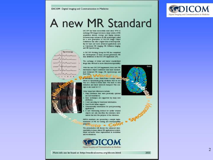
Spectroscopy Color Multi-stack Dimensions Raw Data A new MR Standard (Supplement 49) Multi-frame Real World Values
The basics of Supplement 49 • Support of newest applications by New attributes • Less ambiguity through Stricter definitions and rules • Clear relationships through Reference mechanism • Header size reduction through Multi-frame technique • Context information from Image Type andDimensions • File size flexibility through Concatenations • Functional images with Real World Values • Functional images with PaletteColor LUT information ALL these improve Interoperability
What is really new ? • Multi-Frame module with its Shared and Non-Shared header parts • Dimensions and Dimension Organizations • Coded Object relationships All these provide CONTEXT INFORMATION ! • Further: • Real World Values • Grayscale images with Color information
The Multi-frame properties • Whenever possible, all images of a scan-series shall become frames in one object. • What is common to all frames, shall not be repeated: Elements that do not change vs. elements that change. These have a different place in the object header. • Related elements are defined in functional groups.
Shared Header Pixel data (not to scale) Non-Shared (per-frame) Header From Single Image to Multi-frame N Objects, N Headers HEADER SIZE REDUCTION = less MB, faster transfer N Frames, One Header KV
Fixed Header Dimension data (not to scale) Pixel data (not to scale) Per-frame header From Single-frame to MultiFrame N Objects, N Headers N Frames, One Header
Multi-Frame not only for MR! The Enhanced MR Image and MR Spectroscopy have a multi- frame structure that is also useful for adoption by other modalities.
Dimensions • MR images can have many attributes that change from frame to frame. • When such a change in an attribute is fundamental for the object, this data element can be structured by the creator as part of the Dimension Module. • It supplies the Context Information required for a specialized workstation. • It also provides an explicit display ordering for the frames in the MR object, that can be used by viewing systems without specific MR application knowledge.
Object Relationships Referenced Image Sequence Source Image Sequence
Diffusion Imaging “Diffusion b-values” from 0 to 8000 and an ADC image
Diffusion Tensor Imaging data Reconstructed Fiber Maps in the colors as seen by the creator
time Stored values Real World Value Slope (0040,9225) non perfused stroke area Real World Value Intercept (0040,9224) RW values delayed perfusion Signal time-to-peak map MR Perfusion Imaging Quantitative data with Real World Values
Cardiac Cine Loops Enables automatic multi-slice / multi-phase display, even for standard workstations
Total body Imaging 5 4 3 2 1 1 2 3 4 5 Display the correct image at the correct spot using Stacks and In-stack positions
Functional Brain Imaging • 10-60 slices • all slices measured in one TR • repeated 100-1000 times to get sufficient signal • leading to > 60,000 images in one object Store thousands of images in one object and display them in a consistent way using Multi-frame Header and Dimension Module
Spectroscopy Imaging Relative NAA peak-height Ratio of Choline and Creatinine peaks
Spectroscopy Relations Relation: MR Image Metabolite map Relative NAA peak-height Ratio of Choline and Creatinine peaks
CT Multi-frame • The CT Multi-frame solution addresses the same data-explosion issues as for MR. • Multi-detector CT generates large volumes of images that need to be provided with sufficient and adequate context information. • Multi-frame solutions may provide more interoperability for navigation and processing. • But, 3D processing requires more than a Multi-frame structure.
Multi-modality - Multi-frame • Implementations for processing of the generated volumetric data will largely benefit from multi-modality implementations of the multi-frame concept. • Common structures may lead to common architecture, which in turn will speed up the implementations.
Implementation Roadmap Who should be the first ?
The timing dilemma for the Enhanced MR object • For the MR vendors: • Why implement it, while nobody is ready to use it? • For the Workstation vendors: • Why implement it, when no one is creating it? • For the PACS vendors: • Support it, but how to deal with the mix of workstations that can yes/no receive it ?
The Negotiation scenario Decide to send New object or send Old object Negotiate Negotiate Decide to send New object or Cancel Archive MR Workstation Decide to send New object or send Old object Negotiate
The Partial Conversion scenario Decide to send New object or send Old object Negotiate Negotiate Decide to send New object or Convert object Archive MR Workstation Decide to send New object or send Old object Negotiate
The Full Conversion scenario Decide to send New object or Convert All old objects will be identical Negotiate Negotiate Decide to send New object or Convert object Archive MR Workstation Decide to send New object or Convert Negotiate
When will implementation be required ? • As soon as stability is proven. • As soon as the object definition is tested • As soon as resulting changes are implemented in the standard. • Or….. So, pacing may be needed As soon as possible!
NEMA Members are funding a Demonstration & Test Tool Purpose: • Prove that implementation is possible • Prove the multi-frame header construction • Prove the use of dimensions • Provide a test tool for vendors and other implementers • Validate implemented objects
Tool availability • The tool is available to the funding participants • It is currently under test • The tool will become available in the public domain in 2003 But…No need to wait with implementation
Collective support is needed The benefits of Supplement 49 as described in this presentation will only be visible if and when: • MR scanners (creators) • DICOM workstations (receivers) • PACS systems (intermediaries) will collectively support the new MR DICOM objects.
What should be done? • Vendors of Workstations / PACSneed to prepare for implementation:
What should be done? • Vendors of Workstations / PACS need to prepare for implementation: • Prepare the color pipeline
What should be done? • Vendors of Workstations / PACS need to prepare for implementation: • Prepare the color pipeline • Prepare your databases for large objects
What should be done? • Vendors of Workstations / PACS need to prepare for implementation: • Prepare the color pipeline • Prepare your databases for large objects • Create your real-world values UI
What should be done? • Vendors of Workstations / PACS need to prepare for implementation: • Prepare the color pipeline • Prepare your databases for large objects • Create your real-world values UI • Prepare for Dimensions and Dimension Organizations
What should be done? • Vendors of Workstations / PACS need to prepare for implementation: • Prepare the color pipeline • Prepare your databases for large objects • Create your real-world values UI • Prepare for Dimensions and Dimension Organizations
What should be done? And finally… or first ? Vendors of Workstations / PACS need to decide on the conversion scenario.
Summary • The New DICOM MR Objects: • will enhance interoperability • will increase cross system functionality • will reduce transfer time • Success requires also action by Workstation and PACS implementers.
Further Information More in-depth discussions and presentations about: • Relationships between images • Concatenations • Dimension Organization • Real World Value mapping • Extended Image Type • Color Mapping can be found at : http://medical.nema.org/dicom/presents.html
Acknowledgement and copyright Images for this presentation were provided by: • GE Medical Systems • Philips Medical Systems • Siemens Medical Systems • The slides of this presentation may be quoted if reference and credit to DICOM WG-16 is properly indicated