Figure 6.12
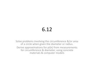
Calcium homeostasis of blood: 9–11 mg/100 ml. BALANCE. BALANCE. Stimulus Falling blood Ca 2+ levels. Thyroid gland. Osteoclasts degrade bone matrix and release Ca 2+ into blood. Parathyroid glands. Parathyroid glands release parathyroid hormone (PTH). PTH. Figure 6.12.
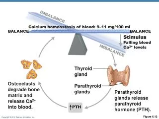

Figure 6.12
E N D
Presentation Transcript
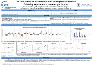
Calcium homeostasis of blood: 9–11 mg/100 ml BALANCE BALANCE Stimulus Falling blood Ca2+ levels Thyroid gland Osteoclasts degrade bone matrix and release Ca2+ into blood. Parathyroid glands Parathyroid glands release parathyroid hormone (PTH). PTH Figure 6.12
Hormonal Control of Blood Ca2+ • May be affected to a lesser extent by calcitonin Blood Ca2+ levels Parafollicular cells of thyroid release calcitonin Osteoblasts deposit calcium salts Blood Ca2+ levels • Leptin has also been shown to influence bone density by inhibiting osteoblasts
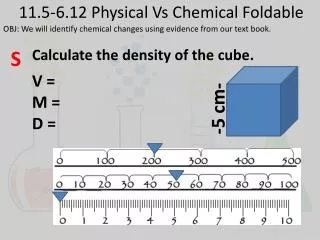
Response to Mechanical Stress • Wolff’s law: A bone grows or remodels in response to forces or demands placed upon it • Observations supporting Wolff’s law: • Handedness (right or left handed) results in bone of one upper limb being thicker and stronger • Curved bones are thickest where they are most likely to buckle • Trabeculae form along lines of stress • Large, bony projections occur where heavy, active muscles attach
Load here (body weight) Head of femur Tension here Compression here Point of no stress Figure 6.13
Classification of Bone Fractures • Bone fractures may be classified by four “either/or” classifications: • Position of bone ends after fracture: • Nondisplaced—ends retain normal position • Displaced—ends out of normal alignment • Completeness of the break • Complete—broken all the way through • Incomplete—not broken all the way through
Classification of Bone Fractures • Orientation of the break to the long axis of the bone: • Linear—parallel to long axis of the bone • Transverse—perpendicular to long axis of the bone • Whether or not the bone ends penetrate the skin: • Compound (open)—bone ends penetrate the skin • Simple (closed)—bone ends do not penetrate the skin
Common Types of Fractures • All fractures can be described in terms of • Location • External appearance • Nature of the break
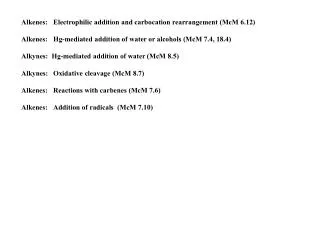
Stages in the Healing of a Bone Fracture • Hematoma forms • Torn blood vessels hemorrhage • Clot (hematoma) forms • Site becomes swollen, painful, and inflamed
Hematoma 1 A hematoma forms. Figure 6.15, step 1
Stages in the Healing of a Bone Fracture • Fibrocartilaginous callus forms • Phagocytic cells clear debris • Osteoblasts begin forming spongy bone within 1 week • Fibroblasts secrete collagen fibers to connect bone ends • Mass of repair tissue now called fibrocartilaginous callus
Externalcallus Internalcallus(fibroustissue andcartilage) Newbloodvessels Spongybonetrabecula 2 Fibrocartilaginouscallus forms. Figure 6.15, step 2
Stages in the Healing of a Bone Fracture • Bony callus formation • New trabeculae form a bony (hard) callus • Bony callus formation continues until firm union is formed in ~2 months
Bonycallus ofspongybone 3 Bony callus forms. Figure 6.15, step 3
Stages in the Healing of a Bone Fracture • Bone remodeling • In response to mechanical stressors over several months • Final structure resembles original
Healedfracture 4 Bone remodelingoccurs. Figure 6.15, step 4
Hematoma Externalcallus Bonycallus ofspongybone Internalcallus(fibroustissue andcartilage) Healedfracture Newbloodvessels Spongybonetrabecula 1 2 3 4 A hematoma forms. Fibrocartilaginouscallus forms. Bony callus forms. Boneremodelingoccurs. Figure 6.15
Homeostatic Imbalances • Osteomalacia and rickets • Calcium salts not deposited • Rickets (childhood disease) causes bowed legs and other bone deformities • Cause: vitamin D deficiency or insufficient dietary calcium
Homeostatic Imbalances • Osteoporosis • Loss of bone mass—bone resorption outpaces deposit • Spongy bone of spine and neck of femur become most susceptible to fracture • Risk factors • Lack of estrogen, calcium or vitamin D; petite body form; immobility; low levels of TSH; diabetes mellitus
Osteoporosis: Treatment and Prevention • Calcium, vitamin D, and fluoride supplements • Weight-bearing exercise throughout life • Hormone (estrogen) replacement therapy (HRT) slows bone loss • Some drugs (Fosamax, SERMs, statins) increase bone mineral density
Paget’s Disease • Excessive and haphazard bone formation and breakdown, usually in spine, pelvis, femur, or skull • Pagetic bone has very high ratio of spongy to compact bone and reduced mineralization • Unknown cause (possibly viral) • Treatment includes calcitonin and biphosphonates
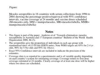
Developmental Aspects of Bones • Embryonic skeleton ossifies predictably so fetal age easily determined from X rays or sonograms • At birth, most long bones are well ossified (except epiphyses)
Parietal bone Occipital bone Frontal bone of skull Mandible Clavicle Scapula Radius Ulna Ribs Humerus Vertebra Ilium Tibia Femur Figure 6.17
Developmental Aspects of Bones • Nearly all bones completely ossified by age 25 • Bone mass decreases with age beginning in 4th decade • Rate of loss determined by genetics and environmental factors • In old age, bone resorption predominates