SEM & FESEM
310 likes | 1.49k Views
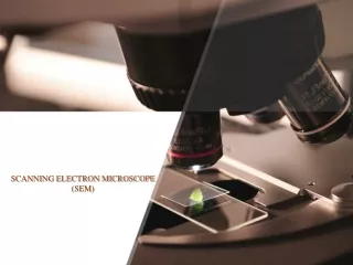
SCANNING ELECTRON MICROSCOPE AND FIELD EMISSION SCANNING ELECTRON MICROSCOPY<br><br>

SEM & FESEM
E N D
Presentation Transcript
Virtual Laboratory Links: https://myscope.training/SEM_simulator.html https://myscope.training/# photogrammetry (using MountainsSEMsoftware)
Virtual Laboratory Links: https://electron-flight-simulator-demo.software.informer.com/download/ Utilizing Monte Carlo Modeling of electron trajectories Electron Flight Simulator is a software tool designed to make your job easier. It can help you understand difficult samples, show the best way to run an analysis, and help explain results to others.
HISTORY • The scanning electron microscope (SEM) was invented by Max Knoll in 1935, at the Telefunken Company in Berlin, for studying the secondary emission properties of television camera tube targets. • The first attempt at building a scanning microscope with a sub-micrometreprobe was made in 1937by Manfred von Ardenne in his private laboratory, also in Berlin. • Further developed by Prof. Sir Charles Oatley and his student Gary Stewart and first time marketed by Cambridge Scientific Instrument Company as the "Stereoscan" in 1965.
What is SEM? SEM = scanning electron microscope A scanning electron microscope (SEM) is a type of electron microscope that produces images of a sample by scanning the surface with a focused beam of electrons. The electrons interact with atoms in the sample, producing various signals that contain information about the surface topography and composition of the sample.
SCANNING ELECTRON MICROSCOPE PRINCIPLE The basic principle is that a beam of electrons is generated by a suitable source. typically a tungsten filament or a field emission gun. The electron beam is accelerated through a high voltage (e.g.: 20 kV) and pass through a system of apertures and electromagnetic lenses to produce a thin beam of electrons. Then the beam scans the surface of the specimen. Electrons are emitted from the specimen by the action of the scanning beam and collected by a suitably-positioned detector.
SCANNING ELECTRON MICROSCOPE • BASIC COMPONENTS • Electronıc console • Electron gun • Electromagnetic lenses • Scanning Coils • Detectors • Sample stage • Vacuum system
BASIC COMPONENTS Electronıc console Focus, Magnification, Brightness, Contrast • Electron gunis used for producing an intense beam of electron. • Thermionic gun thermal energy • Field emission gun electric field • Electromagnetic lenses • LENSESis used to produce clear and Detail images. • Condenser lens reduces the diameter of the electron beam • Objective lens focuses electron beam
BASIC COMPONENTS ScanningCoils After the beam is focused, scanning coils are used to deflect the beam in the X and Y axes so that it scans in a raster fashion over the surface of the sample. Sample stage The container at the end of the column is called the Sample Chamber. The sample stage and the electron detector sit in here. Detectors When the electron beam interacts with a sample in a scanning electron microscope (SEM), multiple events happen. In general, different detectors are needed to distinguish secondary electrons, backscattered electrons, or characteristic x-rays.
Continue….. • VACUUM CHAMBER • SEMs require a vacuum to operate. • Without a vacuum, the electron beam generated by the electron gun would encounter constant interference from air particles in the atmosphere. • Not only would these particles block the path of the electron beam, they would also be knocked out of the air and onto the specimen. which would distort the surface of the specimen.
HOW THE SEM WORKS • The SEM uses electrons instead of light to form an image. • A beam of electrons is produced at the top of the microscope by heating of a metallic filament. • The electron beam follows a vertical path through the column of the microscope. It makes its way through electromagnetic lenses which focus and direct the beam down towards the sample. • Once it hits the sample, other electrons ( backscattered or secondary) are ejected from the sample. • Detectors collect the secondary or backscattered electrons, and convert them to a signal that is sent to a viewing screen similar to the one in an ordinary television, producing an image.
CHEMICAL ANALYSIS! Chemical analysis with a scanning electron microscope and it works like this when the fast electrons of the electron beam the primary electrons reach the surface they knock out electrons of the specimen material these are the secondary electrons used for image formation what happens in detail shows a schematically drawn atom from near the surface just like any other atom it consists of a positively charged nucleus and negatively charged electrons the electrons stay in energetically well defined shells around the nucleus a primary electron comes from above and accidentally knocks out an electron from the K shell of the sample atom a vacant place remains as indicated by the yellow circle this state is unstable an electron from the L shell fills the gap and the energy difference is released in the form of a characteristic x-ray photon this x-ray photon is called characteristic because its energy is quite characteristic or typical for the particular element now another transition follows and finally the atom repairs itself with an electron from the vicinity a free electron so x-ray radiation is generated during the operation of the scanning electron
Continue….. microscope namely X radiation which is characteristic for the chemical elements present in the sample if the energy and the intensity of the radiation are measured with an x-ray detector then the chemical composition of the sample can be determined here the typical x-ray spectrum of the piece of jewelry builds up chemical analysis is then carried out using sophisticated methods with the help of a computer certain limitations must indeed be minded but it's definitely a fine method and above all it is completely non-destructive and even very small spots on a sample can be analyzed and what has happened to the small cobalt plate which was one of the samples it was used to calibrate the measurement this means to adjust it finally the chemical analysis is finished now a piece of jewelry is genuine the gold content amounts to about 70% the balance is silver and copper.
Images Taken by SEM! Scanning electron micrograph of the eggs of a European cabbage butterfly (Pierisrapae).
A video illustrating a typical practical magnification range of a scanning electron microscope designed for biological specimens. The video starts at 25×, about 6 mm across the whole field of view, and zooms in to 12000×, about 12 μm across the whole field of view. The spherical objects are glass beads with a diameter of 10 μm, similar in diameter to a red blood cell.
Applications • SEMs have a variety of applications in a number of scientific and industry-related fields, especially where characterizations of solid materials is beneficial. • In addition to topographical. morphological and compositional information, a Scanning Electron Microscope can detect and analyze surface fractures, provide information in microstructures, examine surface contaminations, reveal spatial variations in chemical compositions, provide qualitative chemical analyses and identify crystalline structures. • In addition, SEMs have practical industrial and technological applications such as semiconductor inspection, production line of miniscule products and assembly of microchips for computers. • SEMs can be as essential research tool in fields such as lift science, biology, gemology, medical and forensic science, metallurgy.
INTRODUCTION • FESEM is the abbreviation of Field Emission Scanning Electron Microscope. A FESEM is microscope that works with electrons (particles with a negative charge) instead of light. These electrons are liberated by a field emission source. The object is scanned by electrons according to a zig-zag pattern. • A FESEM is used to visualize very small topographic details on the surface or entire or fractioned objects. Researchers in biology, chemistry and physics apply this technique to observe structures that may be as small as 1 nanometer (= billion of a millimeter). The FESEM may be employed for example to study organelles and DNA material in cells, synthetically polymers, and coatings on microchips. The microscope that has served as an example for the virtual FESEM is a Jeol6330 that is coupled to a special freeze-fracturing device. • What is difference between Fesem and SEM? • Field emission scanning electron microscopy (FESEM) provides topographical and elemental information at magnifications of 10x to 300,000x, with virtually unlimited depth of field. Compared with convention scanning electron microscopy (SEM), field emission SEM (FESEM) produces clearer, less electrostatically distorted images with spatial resolution down to 1 1/2 nanometers – three to six times better.
CONTINUE….. Overview of the FESEM system
CONTINUE….. • Advantages of FESEM • The ability to examine smaller-area contamination spots at electron accelerating voltages compatible with energy dispersive spectroscopy (EDS). • Reduced penetration of low-kinetic-energy electrons probes closer to the immediate material surface. • High-quality, low-voltage images with negligible electrical charging of samples (accelerating voltages ranging from 0.5 to 30 kilovolts). • Essentially no need for placing conducting coatings on insulating materials. For ultra-high-magnification imaging, we use in-lens FESEM. • Applications of FESEM • Semiconductor device cross section analyses for gate widths, gate oxides, film thicknesses, and construction details • Advanced coating thickness and structure uniformity determination • Small contamination feature geometry and elemental composition measurement