
ATOMIC FORCE MICROSCOPY
E N D
Presentation Transcript
KEY PRINCIPLES There is plenty of room at bottom Micro world 10-6 Physical properties remain same Nano world 10-9 properties totally changed Bulk Material
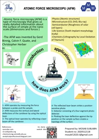
Atomic Force Microscopy(AFM) • The AFM, also known as the atomic force microscope (AFM) is a sort scanner probe. Its principal functions include measuring characteristics like height, magnetism and friction on nanometer fraction. • To investigate non-conductive materials like protein.
Need for AFM • STM was invented in 1981 which gives the image of matter at Nano scale level. • But in STM the sample must be conducting and can work only in vacuum. • So to counter these problems of STM, Gerd Binning invented AFM in 1982 & got Nobel prize for his great invention. • It can make images and manipulation of matter at Nano scale. • It is also called scanning force microscopy, • In simple words we can also say that AFM is the improve version of STM.
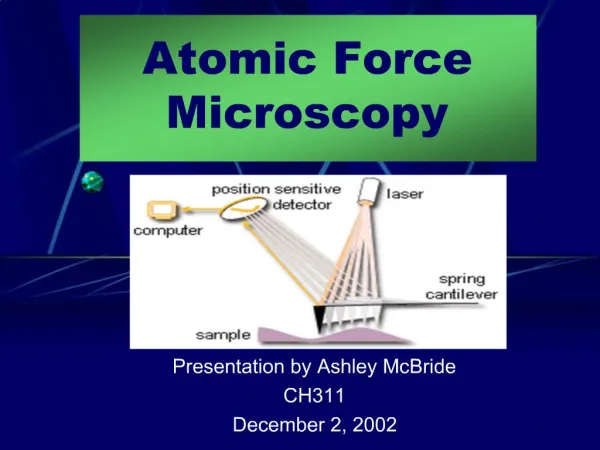
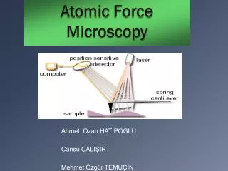
PARTS Cantilever The main component of AFM is Cantilever a metal sheet having a sharp tip at the end. • 2. Tip A very fine tip of size of order of nanometer is composed of Silicon(Si) And is attached at the end of cantilever, Hardly combined of 7 to 8 atoms.
4. Platform The sample under investigation is placed on platform • 5. Piezoelectric tube • The platform containing sample is mounted on the piezoelectric tube Which can move sample in XYZ direction to scanning for 3D view
6. LASER 7. Photoconductor It converts laser light/ energy into electrical energy and then these electric current signals detected by computer it will analyze the current and displayed them on screen To detect the deflection in cantilever To detect the deflection Between the tip atoms and sample atoms
Principle AFM works on the principle that a fine sharp tip of the order of nanometer attached to cantilever when brought into close to a sample surface The Vander walls forces between the sample produces are deflection of cantilever according to the hooks law the reflection is measured by laser spot reflected from the top of cantilever into photo detector. 1.Contact mode e.g. hard metals 2.Non contact mode e.g for light surfaces like biological samples The most preferable working model is non contact mode As in contact mode the are more number of chances of damaging the Cantilever tip. • Modes of working
Advantages • 3D image with high resolution • No need of vaccume like Electron microscope • No need of illumination or focusing • Non conducting sample use • Disadvantages • More scan time • More chances of sample distortion