Download
1 / 48
490 likes | 671 Views
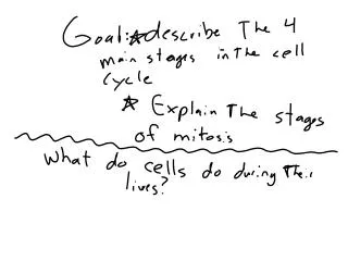
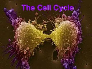
The Cell Cycle and its implications in diseases. Hansjörg Hauser Dept. of Gene Regulation and Differentiation Molecular Biotechnology Helmholtz Centre for Infection Research, Braunschweig. Cell division is a prerequisite for life. Microorganisms reproduce by cell division

E N D
The Cell Cycle and its implications in diseases Hansjörg Hauser Dept. of Gene Regulation and Differentiation Molecular Biotechnology Helmholtz Centre for Infection Research, Braunschweig
Cell division is a prerequisite for life • Microorganisms reproduce by cell division • Mammals need cell division during embryogenesis and for tissue homeostasis Example: Adult humans produce several milions of new cells per second (more than 1011 per day – about 100 grams)
Cell division can be fast or slow • Microorganisms > 20 min per division • Multicellular organisms: 8 min and several weeks per division • All species can halt cell division
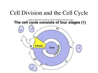
The Cell Cycle M G2 G1 Start, G1 Checkpoint Point of no return S S
Methods to measure cell division • Counting • Amount of DNA • Enzymatic activities • Incorporation of labeled DNA precursors • Cell cycle analysis (FACS) • Dilution of dyes • Time lapse microscopy
While cdks are constitutively expressed the appearance of cyclins in the cell cycle is transient – they cycle The presence of cyclins regulates the activity of the cdks
Cyclic activity of Cyclin kinases Temporal control of the animal cell cycle. The cyclin-E-, cyclin-A- and cyclin-B-dependent kinases are active at different times in the cell cycle. On this basis, cyclin E–Cdk2 appears to have a role in promoting S phase, cyclin A–Cdk2 in S phase and at G2-to-M phase, and cyclin B–Cdk1 during mitosis. Cyclin B1–Cdk1 is activated at the end of G2 phase by the phosphatase Cdc25.
The kinase activity of cdc-cyclin compexes is regulated by phosphorylation and dephosphorylation Examples MO16 is an activating kinase Wee1 is an inhibitory kinase cdc25 is a phosphatase that removes the inhibitory phosphate from the cdk
Regulation of cyclin-dependent kinases. Arrowheads represent activating events and perpendicular ends represent inhibitoryevents. Genes known to perform the indicated functions are listedbelow. Both cyclins and some CKIs (Cdk inhibitors) are regulatedby synthesis and ubiquitin-mediated proteolysis. Checkpoint pathwayscould act to promote inhibitory pathways or inhibit activatingpathways to cause cell cycle arrest
Example: The CKI (cdk inhibitor) p27 p27 inhibits cdk2 and thereby the relvant complexes CyclinE/cdk2 and CyclinA/cdk2 p27 underlies multiple regulations: transcriptional, translational, degradation, localisation (cytoplasmic versus nuclear), phosphorylation, sequestering by binding to CyclinD1/cdk4 (without becoming inactivated)
The progression through the cell cycle underlies many controls: Example DNA replication A re-replication block ensures that no segment of DNA is replicated more than once Passage through mitosis removes the re-replication block Feedback controls generally depend on inhibitory signals
Checkpoint pathways (A) A genetic pathway illustrating intrinsic and extrinsic checkpoint mechanisms. Letters representcell cycle processes. The pathway shown as red symbols indicates an intrinsiccheckpoint mechanism that operates to ensure that event C is completedbefore event E. After event B is completed, an inhibitory signalis activated that blocks completion of event E. After event Cis completed, a signal is sent to turn off the inhibitory signalfrom B, thereby allowing completion of E. The blue symbols representan extrinsic mechanism that is activated when defects such asDNA damage or spindle errors are detected. It is arbitrarily locatedon the D to E pathway but could also function by inhibiting alater step in the B to C pathway. In that case, the extrinsicpathway would utilize the intrinsic mechanism for cell cycle arrest.Mutations in any of the red or blue symbols would result in acheckpoint-effective phenotype.
DNA damage leads to a block in cell cycle progression Replication of damaged DNA would fix mutations for all daughter cells
Possible biochemical function of the Rad24 group of checkpoint proteins. Rad24, together with the four small subunits of RFC, is a component of a pentameric complex. By analogy with RFC, this complex might recognise the transition between ssDNA and dsDNA. Such a structure is produced by many repair pathways but the Rad24 complex may only efficiently recognise it in the context of repair complexes (not shown here). Once the Rad24 complex is bound, it then functions to recruit the ‘PCNA-like’ Rad17/Mec3/Ddc1 complex to the DNA, followed by additional recruitment of checkpoint proteins involved in signal transduction (e.g. Mec1 and Rad53)
Gene expression in G1 G1 S G0 Activation of delayed response genes: E2F Cyclins E, D DNA synthesis genes early response genes: fos, jun,..
Resting cells: The retinoblastoma protein Rb blocks cell cycle progression in G1 by binding to and sequestering E2F Rb-P Rb + E2F Phosphorylation causes Inactivation of Rb Rb : E2F
Target of the CyclinE/cdk2 and CyclinD/cdk4(6) complexes Phosphorylation of pRB
Cell cycle progression by growth factors Phosphorylation causes Inactivation of Rb Proliferation Rb-P Rb E2F CyclinD.cdk4 Rb: E2F CyclinD Rb captures E2F: E2F cannot activate proproliferative genes MAPK pathway Ras EGF
Cell cycle progression Proliferation Growth block Phosphorylation causes Inactivation of Rb Rb captures E2F, so that it cannot activate proproliferative genes Rb-P Rb + E2F CyclinD.cdk4 CyclinD Rb: E2F P16 Ink4A MAPK pathway Ras
Mammalian cells: The protein p53 is sensing DNA damage p53 becomes phosphorylated and stabilized
p53 is a transcriptional activator: One of the genes induced by p53 is p21, an inhibitor of the cdk4 kinase activity p27 Rb captures E2F, so that it cannot activate proproliferative genes CyclinE.cdk2 Rb-P Rb p53 + E2F CyclinD.cdk4 p21 CyclinD Rb: E2F p16 Ink4A MAPK pathway Ras
Cell cycle control in mono- versus multicellular systems • Monocellular systems: • Unlimited proliferation • Control by size, nutrients and sex • Multicellular systems: • Proliferation is limited to specific regions and circumstances: Growth factors, cell:cell-interactions, • In mammals growth and proliferation are independently regulated
Influence of cellular and viral proteins in the cell cycle machinery
Growth factor stimulation through membrane receptors Extracellular ligand binding domain Transmembrane domain Tyrosine kinase domain EGF receptor FGF receptor
Growth factor stimulation through a membrane receptor EGF-receptor Tyrosine kinase
p16 Cycl D:CDK4 p15 Smads p27 RB Cycl E:CDK4 - p21 E2Fs Changes in Gene Expression Cell Proliferation (Cell Cycle) Cell growth inhibitors that act through a membrane receptor Anti-Growth-Factors e.g. TGFß TGFß-R
Rb regulates the cell cycle by binding nuclear transcription factors E2F/myc Rb-P Rb + E2F Phosphorylation causes Inactivation of Rb E2F Rb myc TGFβ
Introduction Current view: • accumulation of multiple mutations within genes of a single cell • mutations confer a competitive advantage for cell growth and (de-) differentiation • mutations lead to initiation and progression of malignancies
Proto-oncogenes • control cell proliferation and differentiation • are expressed in all subcellular compartments (nucleus, cytoplasm, cell surface) • act as protein kinases, growth factors, growth factor receptors, or membrane associated signal transducers
Oncogenes • Mutations in proto-oncogenes alter the normal structure and/or expression pattern • Act in a dominant fashion • gain of function
Mechanisms of oncogene action Biochemically, there are three known mechanisms by which these genes act: • phosphorylation of proteins, with serine, threonine and tyrosine as substrates • signal transmission by GTPases • regulation of DNA transcription
Tumor Suppressor Genes • Have normal, diverse functions to regulate cell growth in a negative fashion (restrain neoplastic growth; act as cellular “brakes”) • physical or functional loss of both alleles frees the cell from constraints imposed by their protein products • loss of function
What causes cancer? • Chemical Carcinogenes • Aflatoxin B1,, Vinylchloride, β-Propiolacton Dimethylsulfate ... • Radiation: • UV, X-Ray, α,-β,-γ-radiation • Viruses • RNA-viruses, DNA-viruses • Spontaneoud mutations Loss of DNA-repair machinery p53
„... manifestation of six essential alterations in cell physiology that collectively dictate malignant growth.“ Cell, Vol. 100, 2000 1. Self-Sufficiency in Growth Signals 2. Insensitivity to Antigrowth Signals 3. Evading Apoptosis 4. Limitless Replicative Potential 5. Sustained Angiogenesis 6. Tissue Invasion and Metastasis
Summary „... manifestation of six essential alterations in cell physiology that collectively dictate malignant growth.“ Cell, Vol. 100, 2000
1. Self-Sufficiency in Growth Signals growth signals (PDGF, TGFα) --> autocrine stimulation overexpression or mutation of receptors (EGF-R, HER2) disruption of intracellular circuits (SOS-Ras-Raf -Map-Kinase)
2. Insensitivity to Antigrowth Signals Disruption of Rb-pathway, downregulation of death receptors
3. Evading Apoptosis Loss of proapoptotic regulators (p53), nonsignaling deathreceptors (FAS)
4. Limitless Replicative Potential Telomerase activation
5. Sustained Angiogenesis Increased expression of angiogen. inducers (VEGF, bFGF) loss of p53 -->downregulation of inhibitors (thrombospondin-1)
6. Tissue Invasion and Metastasis „Out-of-order“ CAMs (E-cadherin), changing integrin expression pattern, overexpression of extracellular proteases, downregulation of protease inhibitor genes