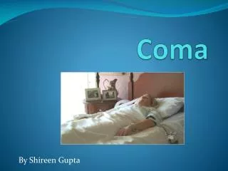
Approach to Coma
Approach to Coma. Dr.Omar Ayoub MBBs, FRCPC Stroke Neurologist and Neurointensivist Assistant Professor of Neurology. Overview. Coma is very broad in all aspects Vague in presentation Vague in approach Long list of differentials Needs very meticulous examination

Approach to Coma
E N D
Presentation Transcript
Approach to Coma Dr.OmarAyoub MBBs, FRCPC Stroke Neurologist and Neurointensivist Assistant Professor of Neurology
Overview • Coma is very broad in all aspects • Vague in presentation • Vague in approach • Long list of differentials • Needs very meticulous examination • Neurological assessment is vital to the diagnosis
Definitions • Confusion: an inability to think with customary speed, clarity, and coherence. • All states of confusion are marked by some degree of inattentiveness and disorientation.
Mild degree: roughly oriented as to time and place, with only occasional irrelevant remarks betraying an incoherence of thinking.
Moderately confused • Carry on a simple conversation for short periods of time, with slow thinking and incoherent, responses are inconsistent, attention span is reduced. • Unable to stay on one topic and to inhibit inappropriate responses. • Usually they are variably disoriented in time and place. • Distractible with any stimulus. • Movements are often tremulous, jerky, and ineffectual. Sequences of movement reveal impersistence
Severely confused • Inattentive persons are usually unable to do more than simplest commands. • Speech is usually limited to a few words or phrases • Disoriented in time and place. • Illusions may lead to fear or agitation. Occasionally, hallucinatory or delusional experiences impart a psychosis.
Drowsiness • Inability to sustain a wakeful state without the application of external stimuli. • Inattentiveness and mild confusion are the rule, both improving with arousal. • The lids droop without closing completely; there may be snoring, the jaw and limb muscles are slack, and the limbs are relaxed.
Stupor • Patient can be roused only by vigorous and repeated stimuli, and does not appear to be unconscious; • Response to spoken commands is either absent or slow and inadequate. • Restless or stereotyped motor activity is common with reduction in the natural shifting of positions. • When left unstimulated, patients quickly drift back into a sleep-like state. • The eyes move outward and upward, a feature that is shared with sleep • Tendon and plantar reflexes and breathing pattern may or may not be altered.
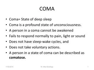
Coma • Incapable of being aroused by external stimuli • Pupillaryreactions, reflex ocular movements, and corneal and brainstem reflexes are preserved in varying degree, muscle tone in the limbs may be increased • Respiration may be slow or rapid, periodic, or deranged in other ways • Vigorous stimulation of the patient or distention of the bladder may cause a stirring or moaning and a quickening of respiration. • These physical signs vary somewhat depending on the cause of coma
Brain death • Complete unresponsiveness to all modes of stimulation, respiratory arrest, and absence of all EEG activity for 24 h. • The central considerations in the diagnosis of brain death are (1) absence of cerebral functions; (2) absence of brainstem functions, including spontaneous respiration (3) irreversibility of the state such as drug overdose.
The Anatomy and Neurophysiology of Alertness and Coma • Paramedian upper brainstem tegmentum and lower diencephalon are the alerting systems of the brain. • The anatomic boundaries of the upper brainstem reticular activating system are the paramedian regions of the upper (rostral) pontine and midbrain tegmentum. • At the thalamic level, it includes the functionally related posterior paramedian, parafascicular, and medial portions of the centromedian and adjacent intralaminar nuclei.
In the brainstem, nuclei of the reticular formation receive collaterals from the spinothalamic and trigeminal-thalamic pathways and project not just to the sensory cortex of the parietal lobe,, but to the whole of the cerebral cortex. • It has become apparent that during wakefulness, there is also a widespread low-voltage fast rhythm (a gamma rhythm that has a frequency of 30 to 60 Hz). This activity, coordinated by the thalamus, has been theorized to synchronize widespread cortical activity and to account perhaps for the unification of modular aspects of experience (color, shape, motion) that are processed in different cortical regions.
Pathologic Anatomy of Coma • Coma is produced by one of two broad groups of problems: • The first is clearly morphologic, consisting either of discrete lesions in the upper brainstem and lower diencephalon (which may be primary or secondary to compression) or of more widespread changes throughout the hemispheres. • The second is metabolic or submicroscopic, resulting in suppression of neuronal activity.
Three pathways • (1) Discernible mass lesion, tumor, abscess, massive edematous infarct, or intracerebral, subarachnoid, subdural, or epidural hemorrhage. • Usually the lesion involves only a portion of the cortex and white matter, leaving much of the cerebrum intact, and it distorts deeper structures. • Cause coma by a lateral displacement of deep central structures, sometimes with herniation of the temporal lobe into the tentorial opening, resulting in compression of the midbrain and subthalamic region of the RAS
(2) A destructive lesion in the thalamus or midbrain, in which case the neurons of the reticular activating are involved directly. • This pathoanatomic pattern characterizes brainstem stroke from basilar artery occlusion, thalamic and upper brainstem hemorrhages, as well as some forms of traumatic damage.
(3) Widespread bilateral damage to the cortex and cerebral white matter as a result of traumatic damage (contusions, diffuse axonal injury), bilateral infarcts or hemorrhages, viral encephalitis, meningitis, hypoxia, or ischemia, as occurs after cardiac arrest. • The coma in these cases results from interruption of thalamocortical impulses or from generalized destruction of cortical neurons.
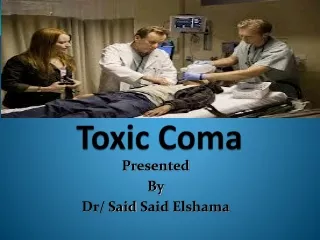
CLINICAL APPROACH TO THE COMATOSE PATIENT • ABC • IF trauma has occurred, one must check for bleeding from a wound or ruptured organ (e.g., spleen or liver), C-spine precaution • Thiamine and glucose • Drug screen and rule out drug intoxication
General Examination • Alterations in vital signs are important aids in diagnosis. • Fever pneumonia or to bacterial meningitis or viral encephalitis. • An excessively high body temperature (42 or 43°C) anticholinergic activity. • Hypothermia is observed in patients with alcoholic or barbiturate intoxication, drowning, exposure to cold, peripheral circulatory failure, and myxedema. • Slow breathing points to opiate or barbiturate intoxication and occasionally to hypothyroidism. • Deep, rapid breathing (Kussmaul respiration) should suggest the presence of pneumonia, diabetic or uremic acidosis, pulmonary edema, or the less common occurrence of an intracranial disease.
Diseases that elevate ICP or damage the brain often cause slow, irregular, or cyclic Cheyne-Stokes respiration. • Vomiting pronounced hypertension, is highly characteristic of cerebral hemorrhage within the hemispheres, brainstem, cerebellum, or subarachnoid space. • The pulse rate, if slow, should suggest heart block from medications such as tricyclic antidepressants or anticonvulsants, or if combined with periodic breathing and hypertension.anincrease in intracranial pressure that reflects the presence of a mass lesion. A myocardial infarction of the inferior wall may also be the cause of bradycardia
Marked hypertension cerebral hemorrhage and hypertensive encephalopathy and sometimes in those with greatly increased intracranial pressure. • Hypotension diabetes, alcohol or barbiturate intoxication, internal hemorrhage, myocardial infarction, dissecting aortic aneurysm, septicemia, Addison disease, or massive brain trauma.
Inspection of the skin • Cyanosis of the lips and nail beds signifies inadequate oxygenation. • Cherry-red coloration is typical of carbon monoxide poisoning. • Multiple bruises (particularly a bruise or boggy area in the scalp), bleeding, CSF leakage from an ear or the nose, or periorbital hemorrhage greatly raises the likelihood of cranial fracture and intracranial trauma. • Telangiectases and hyperemia of the face and conjunctivae of alcoholism; • Myxedema imparts a characteristic puffiness of the face, and hypopituitarism. • Marked pallor suggests internal hemorrhage.
A maculohemorrhagic rash indicates the possibility of meningococcal infection, staphylococcal endocarditis, typhus, or Rocky Mountain fever. • Excessive sweating suggests hypoglycemia or shock, and excessively dry skin, diabetic acidosis or uremia. • Skin turgor is reduced in dehydration. • Large blisters, acute barbiturate, alcohol, and opiate intoxication. • TTP, DIC, and fat embolism diffuse petechiae.
The odor of the breath Alcohol, The spoiled-fruit odor of diabetic coma, the uriniferous odor of uremia, the musty fetor of hepatic coma, and the burnt almond odor of cyanide poisoning.
Neurologic Examination • Grimacing and deft avoidance movements of the stimulated parts are preserved in light coma; their presence substantiates the integrity of corticobulbar and corticospinal tracts. • Yawning and spontaneous shifting of body positions indicate a minimal degree of unresponsiveness. • It is usually possible to determine whether coma is associated with meningeal irritation . • It should be noted that in some patients the signs of meningeal irritation do not develop for 12 to 24 h after the onset of subarachnoid hemorrhage.
Resistance to movement of the neck in all directions may be part of generalized muscular rigidity (as in phenothiazine intoxication) or indicate disease of the cervical spine. • A temporal lobe or cerebellarherniation or decerebrate rigidity may also limit passive flexion of the neck and be confused with meningeal irritation. • A moan or grimace may be provoked by painful stimuli on one side but not on the other, reflecting the presence of a hemianesthesia; also during grimacing, facial weakness may be noted.
Pupillary Reactions • A unilaterally enlarged pupil (5.5 mm diameter) is an early indicator of stretching or compression of the third nerve and reflects the presence of an ipsilateral hemispheral mass. • A loss of light reaction alone usually precedes enlargement of the pupil. • As a transitional phenomenon, the pupil may become oval or pear-shaped or appear to be off center (corectopia) due to a differential loss of innervation of a portion of the pupillary sphincter.
The light-unreactive pupil continues to enlarge to a size of 6 to 9 mm diameter and is soon joined by a slight outward deviation of the eye, • As midbrain displacement continues, both pupils dilate and become unreactive to light as a result of compression of the oculomotor nuclei in the rostral midbrain • Pontine tegmental lesions cause extremely miotic pupils (<1 mm in diameter) • The ipsilateralpupillary dilatation from pinching the side of the neck (the ciliospinal reflex) is also lost in brainstem lesions.
A Horner syndrome (miosis, ptosis, and reduced facial sweating) may be observed ipsilateral to a lesion of brainstem or hypothalamus or as a sign of dissection of ICA. • With coma due to drug intoxications and metabolic disorders, pupillary reactions are usually spared, but there are notable exceptions. • Opiates or barbiturates cause pinpoint pupils with a constriction to light • Poisoning with atropine or with drugs that have atropinic qualities, especially the tricyclic antidepressants (wide dilation and fixity of pupils)
Movements of Eyes and Eyelids and Corneal Responses • The eyes may be turned down and inward (looking at the nose) with hematomas or ischemic lesions of the thalamus and upper midbrain (a variant of Parinaudsyndrome) • Retraction and convergence nystagmus and "ocular bobbing," occur with lesions in the tegmentum of the midbrain and pons, respectively. • Ocular dipping in which the eyes move down slowly and return rapidly to the meridian, may be observed with coma due to anoxia and drug intoxications; horizontal eye movements are preserved
Oculovestibular or caloric test • Irrigation of each ear with 10 mL of cold water (or room-temperature water if the patient is not comatose) normally causes slow conjugate deviation of the eyes toward the irrigated ear, followed in a few seconds by compensatory nystagmus (fast component away from the stimulated side). • The ears are irrigated separately several minutes apart. • In comatose patients, the fast "corrective" phase of nystagmus is lost and the eyes are tonically deflected to the side irrigated with cold water or away from the side irrigated with warm water; this position may be held for 2 to 3 min.
With brainstem lesions, these vestibulo-ocular reflexes are lost or disrupted. • If only one eye abducts and the other fails to adduct, one can conclude that the medial longitudinal fasciculus has been interrupted (on the side of adductor paralysis). • The opposite, abducens palsy, is indicated by an esotropic resting position and a lack of outward deviation of one eye with the reflex maneuvers. • The complete absence of ocular movement in response to oculovestibular testing indicates a severe disruption of brainstem tegmental pathways in the pons or midbrain or, as mentioned, a profound overdose of sedative or anesthetic drugs.
Posturing in the Comatose Patient • Decerebrate rigidity, which in its fully developed form consists of opisthotonos, clenching of the jaws, and stiff extension of the limbs, with internal rotation of the arms and plantar flexion of the feet (brainstem at the intercollicular level). • Decorticate rigidity, with arm or arms in flexion and adduction and leg(s) extended, signifies lesions at a higher level, in cerebral white matter or IC and thalamus.