Download
1 / 86
880 likes | 1.13k Views
38. Circulatory Systems. Chapter 38 Circulatory Systems. Key Concepts 38.1 Circulatory Systems Can Be Open or Closed 38.2 Circulatory Systems May Have Separate Pulmonary and Systemic Circuits 38.3 A Beating Heart Propels the Blood 38.4 Blood Consists of Cells Suspended in Plasma.

E N D
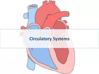
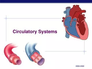
38 Circulatory Systems
Chapter 38 Circulatory Systems • Key Concepts • 38.1 Circulatory Systems Can Be Open or Closed • 38.2 Circulatory Systems May Have Separate Pulmonary and Systemic Circuits • 38.3 A Beating Heart Propels the Blood • 38.4 Blood Consists of Cells Suspended in Plasma
Chapter 38 Circulatory Systems • Key Concepts • 38.5 Blood Circulates through Arteries, Capillaries, and Veins • 38.6 Circulation Is Regulated by Autoregulation, Nerves, and Hormones
Chapter 38 Opening Question What are the critical factors that determine whether a person recovers from a heart attack?
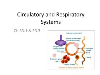
Concept 38.1 Circulatory Systems Can Be Open or Closed • The function of a circulatory system is to transport substances around the body. • It consists of: • Muscular pump—theheart • Fluid—blood • Series of conduits—blood vessels
Concept 38.1 Circulatory Systems Can Be Open or Closed • Some animals do not need circulatory systems: • Single-celled organisms exchange directly with the environment • Structures and body shapes allow exchange between cells and the environment • Gastrovascular systems bring the external environment inside the animal
Concept 38.1 Circulatory Systems Can Be Open or Closed • Open circulatory system: • The circulatory fluid—hemolymph—leaves circulatory system and moves between cells and tissues • Flows back into heart or circulatory vessels • Opencirculatory systems are found in arthropods and mollusks.
Concept 38.1 Circulatory Systems Can Be Open or Closed • Closed circulatory system—blood vessels keep circulatory fluid (blood) separate from the fluid around cells (interstitial fluid). • Blood consists of liquid blood plasma and blood cells. • Water and small molecules leak out through capillaries—blood plasma and interstitial fluid make up extracellular fluid.
Concept 38.1 Circulatory Systems Can Be Open or Closed • The closed vascular system contains: • Arteries—carry blood away from the heart and branch into arterioles that feed the capillary beds • Capillaries—the site of exchange between blood and interstitial fluid • Venules—drain the capillary beds and form veins, which deliver blood back to the heart
Concept 38.1 Circulatory Systems Can Be Open or Closed • Advantages of closed circulatory systems: • Circulatory fluid can flow more rapidly • Blood flow to specific tissues can be controlled by varying resistance • Specialized cells and molecules that transport oxygen, hormones, and nutrients can be kept in the vessels
Concept 38.2 Circulatory Systems May Have Separate Pulmonary and Systemic Circuits • In fish, there is a single circuit; in birds, crocodiles and mammals, there are separate circuits: • Pulmonary circuit—blood is pumped from the heart to the lungs and back again • Systemic circuit—blood travels from the heart to the rest of the body and back to the heart
Concept 38.2 Circulatory Systems May Have Separate Pulmonary and Systemic Circuits • Fish hearts have two chambers: • Atrium—receives blood from the body • Ventricle—receives pumped blood from the atrium and sends it to the gills, arranged on gill arches • Blood flows through afferent arterioles into the gill arch. • Blood leaves in efferent arterioles, which join together in the aorta.
Concept 38.2 Circulatory Systems May Have Separate Pulmonary and Systemic Circuits • Lungfish have adapted to breathe in air as well as in water. • A lung formed from the gut functions in air. • A divided atrium separates blood into pulmonary and systemic circuits—it can receive blood from either the lung or other tissues.
Concept 38.2 Circulatory Systems May Have Separate Pulmonary and Systemic Circuits • Amphibians have three-chambered hearts. • A ventricle pumps blood to the lungs and body. • One atrium receives oxygenated blood from the lungs, a second atrium receives blood from the body. • The ventricle directs the flow to the pulmonary or systemic circuit.
Concept 38.2 Circulatory Systems May Have Separate Pulmonary and Systemic Circuits • Reptiles have three- or four-chambered hearts and two aortas: • The left aorta receives oxygenated blood from the left side of the ventricle and delivers it to the body • The right aorta can receive blood from either side of the ventricle
Concept 38.2 Circulatory Systems May Have Separate Pulmonary and Systemic Circuits • The reptilian ventricle is partly divided by a septum. • When the animal is breathing, blood flows to the pulmonary circuit. • When the animal is not breathing, blood flows to the systemic circuit.
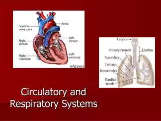
Concept 38.2 Circulatory Systems May Have Separate Pulmonary and Systemic Circuits • Crocodilians, birds, and mammals have four-chambered hearts and separate pulmonary and systemic circuits. • Deoxygenated blood from the body arrives at the right atrium and flows into the right ventricle. • Right ventricle pumps blood through pulmonary arteries to lungs to pick up oxygen—then back to the left atrium of the heart through pulmonary arteries.
Concept 38.2 Circulatory Systems May Have Separate Pulmonary and Systemic Circuits • Oxygenated blood flows from the left atrium into the left ventricle—it contracts and sends blood through the aorta to the rest of the body. • Oxygen-depleted blood returns to the right atrium through the vena cavae, large veins.
Concept 38.2 Circulatory Systems May Have Separate Pulmonary and Systemic Circuits • Separate circuits have advantages: • Oxygenated blood can be distributed at higher pressure and flow than is possible in fishes. • Blood in each system cannot mix—systemic circuit always receives blood with higher O2 content. • Circuits can operate at different pressures.
Concept 38.3 A Beating Heart Propels the Blood • The human heart has four chambers—two atria and two ventricles. • The right atrium receives oxygen-depleted blood—it then flows through an atrioventricular (AV) valve into the right ventricle. • When the right ventricle contracts, the flaps of the AV valve close, to prevent backflow. • Blood is pumped through pulmonary artery to the lungs and pulmonary valve closes.
Concept 38.3 A Beating Heart Propels the Blood • Oxygenated blood returns via pulmonary veins to the left atrium. • Blood flows into left ventricle through another AV valve. • Left ventricle contracts forcefully to send blood through the aorta—then relaxes. • The aortic valve at base of aorta then closes, to prevent backflow.
Concept 38.3 A Beating Heart Propels the Blood • The ventricles can adjust the force of their contractions to meet demands of exercise. • The Frank-Starling law is a property of cardiac muscle cells: • When they are stretched, as occurs when returning blood volume increases, they contract more forcefully.
Concept 38.3 A Beating Heart Propels the Blood • The cycle of cardiac contraction and relaxation is the cardiac cycle. • Two phases: • Systole—when ventricles contract • Diastole—when ventricles relax • The atria contract just before the ventricles, to add blood volume to the ventricles. • Heart murmurs are sounds made by valves that do not close completely.
Concept 38.3 A Beating Heart Propels the Blood • Cardiac muscle functions as a pump: • Cells are in electrical contact with each other through gap junctions—spread of action potentials stimulates contraction in unison. • Some cells are pacemaker cells and can initiate action potentials without input from the nervous system. • The primary pacemaker cells are in the sinoatrial node.
Concept 38.3 A Beating Heart Propels the Blood • Action potentials in pacemaker cells are generated by voltage-gated Ca2+ channels. • The resting membrane potential of these cells is not stable and gradually drifts upward.
Concept 38.3 A Beating Heart Propels the Blood • Ion channels in pacemaker cells are different from other cardiac cells: • Na+ channels are more permeable to sodium influx, so resting potential is higher. • K+ channels that open after action potential to repolarize cell eventually close—K+ inside cell causes membrane potential to drift upwards toward threshold
Concept 38.3 A Beating Heart Propels the Blood • Pacemaker cells initiate contractions—the heart does not need nerve signals to beat. • The nervous system controls heart rate by influencing resting potential: • Norepinephrine from sympathetic nerves increases permeability of Na+/K+ and Ca2+ channels. • The resting potential rises more quickly and action potentials are closer together.
Concept 38.3 A Beating Heart Propels the Blood • Opposite effect from parasympathetic nerves: • Acetylcholine increases permeability of K+ and decreases that of Ca2+ channels. • The resting potential rises more slowly and action potentials are farther apart.
Figure 38.4 The Autonomic Nervous System Controls Heart Rate (Part 1)
Figure 38.4 The Autonomic Nervous System Controls Heart Rate (Part 2)
Concept 38.3 A Beating Heart Propels the Blood • Heart muscle contraction is coordinated. • An action potential is generated in the sinoatrial node. • The action potential spreads through gap junctions in the atria and they contract together, but it does not spread to the ventricles.
Concept 38.3 A Beating Heart Propels the Blood • The action potential in the atria stimulates the atrioventricular node. • The node consists of non-contracting cells that send action potentials to the ventricles via the bundle of His. • The bundle divides into right and left bundle branches that run to the tips of the ventricles and then spread throughout—called Purkinje fibers. • A contraction spreads rapidly and evenly throughout the ventricles.
Concept 38.3 A Beating Heart Propels the Blood • Ventricular muscle fibers contract for much longer than skeletal muscle fibers. • Their extended action potential is due to a longer opening of voltage-gated Ca2+ channels and increased availability of Ca2+ to stimulate contraction.
Concept 38.3 A Beating Heart Propels the Blood • An electrocardiogram (ECG or EKG) uses electrodes to record events in the cardiac cycle. • Large action potentials in the heart cause electrical current to flow outward to all parts of the body. • Electrodes register the voltage difference at different times. • Wave patterns of an ECG are labeled by letters P, Q, R, S, and T—each representing an event