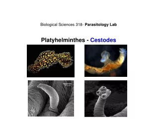

Cestodes
E N D
Presentation Transcript
Cestodes Taeniasaginata Taeniasolium
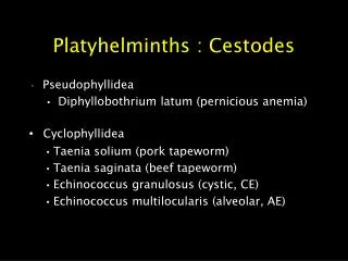
Classification • Cestodes belong to Phylum –Platyhelminthis Class—Cestoidea Subclass—Eucestoda Order—Pseudophyllidea Cyclophyllidea
Classification based on Habitat • Intestinal Cestodes—Diphyllobotrium , Taenia,Hymenolepis,Dipylidiumspicies • Somatic or Tissue Cestodes(Larvae in Human Muscle or Organ)—T.solium,T.multiceps,Echinococcus,Spirometra species
Order Family Genus PseudophyllideaDiphyllobothriidaeDiphyllobdthrium Spirometra CyclophyllideaTaeniidaeTaenia Echinococcus HymenolepididaeHymenolepis DipylidiidaeDipyllidium
They exist in three morphology—Adult worm , Egg and, Larva . • Adult worm---intestine of human and animals . They are usually long, segmented, Flattened and tape like. • Few mm to meters—1 to 4cm to 10 mts.
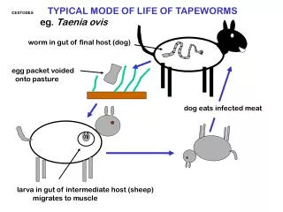
HOSTS • INTERMEDIATE: harbors the immature forms of the parasite. Insect of herbivorous vertebrate that ingest parasite eggs • DEFINITIVE: Harbors the mature forms of the parasite. Carnivorous or omnivorous mammal that acquires infection by consuming larval cysts in the uncooked tissues of an IH
scolex neck strobila Tapeworms • The body plan of adult cestodes includes a scolex (looks like the “head”), a neck and strobila that can extend for only a few proglottids or thousands • The strobila is not truly metameric though as several organs like the excretory system extend through the entire worm • Proglottid: each individual segment • Most worms are very long: occupying the entire length of small intestine
Taeniasaginata (Beef tapeworm) • Ranges in length from 6-30 ft • Geographic distribution: cosmopolitan. • Most common where poor sanitation and no inspection of meat combine • Africa and South America • Transmission: Ingestion of larval form in undercooked beef • In N. America 1 in 100 is infected. In third-world nations could be up to 10% • No symptoms or some abdominal discomfort • Diagnosis: finding eggs or proglottids in feces
Taeniasolium • T. solium has a scolex (A) with four suckers and a double crown of hooks, a narrow neck, and a large strobila (2-4 m) (B) consisting of several hundred proglottids. • About 2 months after ingestion, proglottids begin to detach from the distal end and are excreted in the feces. • Each segment contains 50-60,000 fertile eggs.
TAENIA SOLIUM • The larval stage of the pork tapeworm infects the human nervous system causing neurocysticercosis. • One of the main causes of epileptic seizures. • Endemic in less developed countries where pigs are raised as food source. Latin America, most of Asia, sub-saharan Africa, and parts of Oceania. The Lancet (2003) 361: 547
Cysticeruscellulosae • Larval stage of T.soliumin tissues • Humans acquire cysticercosis through faecal-oral contamination with T. solium eggs • The oncosphere in the eggs is released by the action of gastric acid and intestinal fluids • Cross the gut wall and enter the bloodstream • They are carried to the muscles and other tissues • They encyst as cysticerciat small terminal vessels (1 cm) (A) and (B) • Neurocysticercosis and ophtalmiccysticercosis serious
Neurocysticercosis • The parasite infects the CNS • Epileptic seizures (58-80% when parenchymal brain cysts). • Intracranial hypertension, hydrocephalus, or both. This syndrome is related to the location of parasites in the cerebral ventricles or vasal cisterns. • Occasionally a cyst may grow larger (giant cyst) • Geographical variation in clinical manifestations
Cysticercusbovis • Larval forms of T.saginata in tissues • Occurs in cattle but not in humans
Lab diagnosis - Intestinal taeniasis • Stool examination • Segments of adult worm • eggs
Lab diagnosis of cysticercosis • Serologic diagnosis: • Antibody assays for cysticercosis: 8 kDa antigens, GP50, FAST-ELISA with the 8 kDA antigen • Antigen-detection assays: circulating antigens means live parasites. Ongoing viable infection. Monoclonal antibodies seem to detect AGs in CSF. • Antibody assays for taeniasis: TSE33 and TSE38 were recognized by a panel of taeniasis but not cysticercocis, patient serum samples.
Neuroimaging diagnosis: CT and MRI provide objective evidence on number and location of cysticerci. Also their viability and the severity of the host inflammatory reaction.
Treatment • Treatment should be individualized based on cyst location, level of immflamation and clinical presentation • Parenchymalcysticercosis with viable cysts: Albendazole 15 (mg/kg/day) with dexamethasone (0.1 mg/kg/day). Praziquantel. • Subarachnoid ccs: antiparasitic therapy • No reason to use antiparasitic drugs to treat dead calcified cysts. Symptomatic therapy. • Surgical therapy: ventricular shunting to resolve hydrocephalus. Also excision of giant cysts or intraventricular cysts • Taeniasis treatment: niclosamide and praziquantel