Download
1 / 12
160 likes | 225 Views
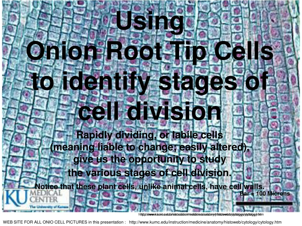
http://www.kumc.edu/instruction/medicine/anatomy/histoweb/cytology/cytology.htm. Bar = 100 Microns. http://www.kumc.edu/instruction/medicine/anatomy/histoweb/cytology/cytology.htm. Using Onion Root Tip Cells to identify stages of cell division

E N D
http://www.kumc.edu/instruction/medicine/anatomy/histoweb/cytology/cytology.htmhttp://www.kumc.edu/instruction/medicine/anatomy/histoweb/cytology/cytology.htm Bar = 100 Microns http://www.kumc.edu/instruction/medicine/anatomy/histoweb/cytology/cytology.htm Using Onion Root Tip Cells to identify stages of cell division Rapidly dividing, or labile cells (meaning liable to change; easily altered), give us the opportunity to study the various stages of cell division. Notice that these plant cells, unlike animal cells, have cell walls. WEB SITE FOR ALL ONIO CELL PICTURES in this presentation : http://www.kumc.edu/instruction/medicine/anatomy/histoweb/cytology/cytology.htm
Bar = 50 Microns Slide 2 Onion Root Tip You will be attempting to find cells in various stages of cell division. Each colored arrow points to chromosomes during one of the stages of division http://www.kumc.edu/instruction/medicine/anatomy/histoweb/cytology/cytology.htm
Slide 3 Onion Root TipYOU WILL BE USING THE MEDIUM POWER OBJECTIVE FIRST. Find a cell (or cells) that look similar to those that which the GREEN arrow is pointing. When you find one, use the microscope arrow (move the slide if/when necessary)to POINT TO THE CELL and SHOW IT TO YOUR TEACHER. After you have had your cell approved, change to HIGH POWERand then draw the individual cell on your paper. Bar = 30 Microns
Slide 3 Onion Root TipIf you were not able to locate this cell on medium power, switch to HIGH POWER. Again, attempt to find a cell that look similar to that which the GREEN arrow is pointing. Once you locate it, use the microscope’s arrow (move the slide if / when necessary) so that it points to the correct cell. REMEMBER YOU MUST SHOW THE CELL to your teacher ! After you have had your located cell approved, draw the individual cell on your paper.
Bar = 30 Microns http://www.kumc.edu/instruction/medicine/anatomy/histoweb/cytology/cytology.htm Slide 4 Onion Root TipYOU WILL BE USING THE MEDIUM POWER OBJECTIVE FIRST. Find a cell (or cells) that look similar to those that which the BLUE arrow is pointing. When you find one, use the microscope arrow (move the slide if/when necessary)to POINT TO THE CELL and SHOW IT TO YOUR TEACHER. After you have had your cell approved, change to HIGH POWERand then draw the individual cell on your paper.
Slide 4 Onion Root TipIf you were not able to locate this cell on medium power, switch to HIGH POWER. Again, attempt to find a cell that look similar to that which the BLUE arrow is pointing. Once you locate it, use the microscope’s arrow (move the slide if / when necessary) so that it points to the correct cell. REMEMBER YOU MUST SHOW THE CELL to your teacher ! After you have had your located cell approved, draw the individual cell on your paper.
Bar = 30 Microns http://www.kumc.edu/instruction/medicine/anatomy/histoweb/cytology/cytology.htm Slide 5 Onion Root TipYOU WILL BE USING THE MEDIUM POWER OBJECTIVE FIRST. Find a cell (or cells) that look similar to those that which the RED arrow is pointing BE SURE THAT YOU SEE THE CHROMOSOMES FORMING INTO 2 CELLS. When you find one, use the microscope arrow (move the slide if/when necessary)to POINT TO THE CELL and SHOW IT TO YOUR TEACHER. After you have had your cell approved, change to HIGH POWERand then draw the individual cell on your paper.
Slide 5 Onion Root TipIf you were not able to locate this cell on medium power, switch to HIGH POWER. Again, attempt to find a cell that look similar to that which the RED arrow is pointing. BE SURE THAT YOU SEE THE CHROMOSOMES FORMING INTO 2 CELLS Once you locate it, use the microscope’s arrow (move the slide if / when necessary) so that it points to the correct cell. REMEMBER YOU MUST SHOW THE CELL to your teacher ! After you have had your located cell approved, draw the individual cell on your paper.
Bar = 30 Microns Slide 6 Onion Root TipYOU WILL BE USING THE MEDIUM POWER OBJECTIVE FIRST. Find a cell (or cells) that look similar to those that which the CIRCLED BLACK arrow is pointing. When you find one, use the microscope arrow (move the slide if/when necessary)to POINT TO THE CELL and SHOW IT TO YOUR TEACHER. After you have had your cell approved, change to HIGH POWERand then draw the individual cell on your paper.
Slide 6 Onion Root TipIf you were not able to locate this cell on medium power, switch to HIGH POWER. Again, attempt to find a cell that look similar to that which the CIRCLED BLACK arrow is pointing. Once you locate it, use the microscope’s arrow (move the slide if / when necessary) so that it points to the correct cell. REMEMBER YOU MUST SHOW THE CELL to your teacher ! After you have had your located cell approved, draw the individual cell on your paper.
WEBSITES • Website for pictures in this presentationhttp://www.kumc.edu/instruction/medicine/anatomy/histoweb/cytology/cytology.htm • Interactive Mitosis Website for Onion Root tip Lab http://bio.rutgers.edu/~gb101/lab2_mitosis/notebook.html
Additional Image Sites • http://www.wellcome.ac.uk/Education-resources/Education-and-learning/Big-Picture/All-issues/The-Cell/Image-galleries-Aspects-of-imaging/WTDV030896.htm • http://biology.about.com/od/mitosis/ig/Mitosis-Image-Gallery/Early-Prophase.htm • http://www.youtube.com/watch?feature=player_embedded&v=NR0mdDJMHIQ