Inelastic x-ray scattering (IXS)
Inelastic x-ray scattering (IXS). Beamline Development Team: Y. Cai , S. Antonelli, D.S . Coburn, A. Cunsolo, J . Keister, C.N . Kodituwakku, D. Levy, L. Reffi, Y . Stetsko, A . Suvorov, Photon Sciences, Brookhaven National Laboratory

Inelastic x-ray scattering (IXS)
E N D
Presentation Transcript
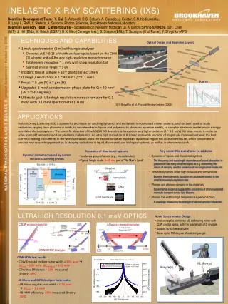
Inelastic x-ray scattering (IXS) Beamline Development Team: Y. Cai, S. Antonelli, D.S. Coburn, A. Cunsolo, J. Keister, C.N. Kodituwakku, D. Levy, L. Reffi, Y. Stetsko, A. Suvorov, Photon Sciences, Brookhaven National Laboratory Beamline Advisory Team:Clement Burns– Spokesperson (Western Michigan U), A. Baron (SPring-8/RIKEN), S.H. Chen (MIT). J. Hill (BNL), M. Krisch (ESRF), H.K. Mao (Carnegie Inst.), S. Shapiro (BNL), T. Scopigno (U of Rome), Y. Shvyd’ko (APS) TECHNIQUES AND CAPABILITIES Optical Design and Beamline Layout • 1 meV spectrometer (5 m) with single analyzer • Operates at E ~ 9.13 keV with analyzer optics based on the CDW [1] scheme and a 4-Bounce high-resolution monochromator • Total energy resolution ~ 1 meV with sharp resolution tail • Scanned energy range: ~ 1 eV • Incident flux at sample > 1010 photons/sec/1meV • Q range / resolution: 0.1 ~ 40 nm-1 / ~ 0.1 nm-1 • Focus: ~ 5 µm (V) x 7 µm (H) • Upgraded 1-meV spectrometer: phase plate for Q > 40 nm-1 (2θ > ~50 degrees) • Ultimate goal: Ultrahigh resolution monochromator for 0.1 meV, with 0.1 meV spectrometer (10 m) 0.1-meV development spectrometer 1-meV spectrometer Size (nm) Source Hemoglobin DNA Lipid membrane [1] Y. Shvyd’ko et al, Physical Review Letters (2006) Applications Inelastic X-ray Scattering (IXS) is a powerful technique for studying dynamics and excitations in condensed matter systems, and has been used to study excitations ranging from phonons in solids, to sound modes in liquids and polymers, to plasmons in simple metals, to complex electronic excitations in strongly correlated electron systems. The scientific objective of the NSLS-II IXS Beamline is focused on very high-resolution (1 ~ 0.1 meV) IXS experiments in order to solve some of the most important problems in dynamics. An ultra high resolution of 0.1 meV represents an order of magnitude improvement over the best currently existing instruments in the world and would allow the exploration of an important dynamical region not accessible thus far, which is expected to provide new research opportunities in studying excitations in liquid, disordered, and biological systems, as well as in phonon research. Dynamic domains covered by current inelastic scattering probes • Key scientific questions to address • Dynamics of liquids and disordered systems • The frequency and wavelength dependence of sound absorption in glasses still has many unsolved topics as e.g. concerning the nature of damping and the existence of non longitudinal modes etc. • Vibration dynamics under high pressure and temperature • Extreme thermodynamic condition are accessible thanks to the small transversal x-ray beam size • Phonon and phonon-damping in bio-materials • Experimental evidence suggests the occurrence of phonon-assisted molecule transport across lipid bilayers • Phonon line width in high temperature superconductors • A challenge: measuring the strength of electron-phonon interaction • Dynamics of disordered systems • Involves a group of atoms (e.g., bio-molecules) • Typical length scale: 5~50 nm, part of “No Man’s Land” Ultrahigh resolution 0.1 meV optics • Novel Spectrometer Design • Analyzer optics combines ML collimating mirror with CDW crystal optics, with 1m total length of D crystals • Support up to five analyzers • Cover up to 135 degree of scattering angle. CDDW monochromator 4-Bounce monochromator CDW/CDDW Analyzer • CDW-CDW test results • CDW-D crystal rocking curve width = 3.65 µrad ΔEtotal = 0.87 meV, ΔEindividual= 0.62 meV • CDW-Ana Efficiency: ~20% measured (theory: 38%) ML Mirror(s) Analyzer(s) • 4B Mono and CDW Analyzer test results • 4B Mono angular scan width = 0.32 µrad ΔEtotal = 1.1 meV • 4B HRM efficiency: ~30% measured (theory: 35%)