Download
1 / 57
570 likes | 824 Views
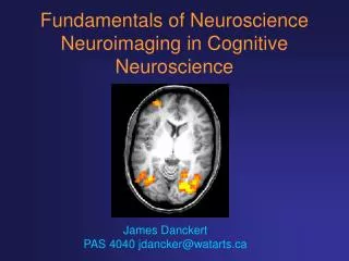
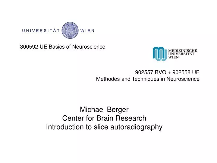
300592 UE Basics of Neuroscience. 902557 BVO + 902558 UE Methodes and Techniques in Neuroscience. Michael Berger Center for Brain Research Introduction to slice autoradiography.

E N D
300592 UE Basics of Neuroscience 902557 BVO + 902558 UE Methodes and Techniques in Neuroscience Michael Berger Center for Brain Research Introduction to slice autoradiography
For binding experiments, most commonly membrane preparations are used (membranes of homogenized tissue). A more ambitious, but also more time-consuming technique is the binding of radioligands to slices of intact tissue, with preserved tissue integrity. Sometimes, this technique is still described as new, but ...
For binding experiments, most commonly membrane preparations are used (membranes of homogenized tissue). A more ambitious, but also more time-consuming technique is the binding of radioligands to slices of intact tissue, with preserved tissue integrity. Sometimes, this technique is still described as new, but ... W.S. Young & M.J. Kuhar (1979) A new method for receptor autoradiography: [3H]opioid receptors in rat brain. Brain Res. 179: 255-270
For binding experiments, most commonly membrane preparations are used (membranes of homogenized tissue). A more ambitious, but also more time-consuming technique is the binding of radioligands to slices of intact tissue, with preserved tissue integrity. Sometimes, this technique is still described as new, but ... W.S. Young & M.J. Kuhar (1979) A new method for receptor autoradiography: [3H]opioid receptors in rat brain. Brain Res. 179: 255-270 ... it‘s probably older than most of you.
Slice-autoradiography allows the semi-microscopic evaluation of binding site distribution in fixed, partially fixed, or unfixed (native) tissue. “Semi-microscopic“ means a resolution down to 50 – 10 µm, i.e. down to cellular dimensions (in mammalian tissue).
Since long, tissue slices have been obtained in good quality from fixed material, i.e. from biological tissue treated with a fixative as formaldehyde. With these fixation techniques, subcellular structures have been visualized since the times of Golgi and Cajal.
Since long, tissue slices have been obtained in good quality from fixed material, i.e. from biological tissue treated with a fixative as formaldehyde. With these fixation techniques, subcellular structures have been visualized since the times of Golgi and Cajal. After these treatments, however, most receptors are no longer recognized by their natural ligands.
For receptor autoradiography, the tissue is only slightly fixed, or not fixed at all. To allow the preparation of thin slices, the tissue is frozen, and kept frozen in a cryostat.
Don‘t freeze the tissue by simply putting it into a – 20 °C refrigerator.
Don‘t freeze the tissue by simply putting it into a – 20 °C refrigerator. (It will look like this – freezing artefacts)
Best tissue quality needs shock-freezing. Here the advice is: shock-freeze to -18 °C. But we go further...
Method of choice: freezing mixture with dry ice and organic solvent (mostly isopentane), kept at – 45 to – 40 °C.
Method of choice: freezing mixture with dry ice and organic solvent (mostly isopentane), kept at – 45 to – 40 °C. Temperature must be supervised with a thermometer: If you wait too long, it will reach – 78 °C (the sublimation temperature of CO2).
To avoid freezing artefacts in the middle of the tissue, at least one dimension must be below 10 mm. Specimens, that are successfully frozen: For a rat brain, a 100 ml beaker will be sufficient.
To avoid freezing artefacts in the middle of the tissue, at least one dimension must be below 10 mm. Specimens, that are successfully frozen: For a rat brain, a 100 ml beaker will be sufficient. For a human brain slice, a 1.000 ml jar will be necessary.
The frozen tissue is transferred to the cryostat chamber (kept at – 10 to – 20 °C) and mounted with...
The frozen tissue is transferred to the cryostat chamber (kept at – 10 to – 20 °C) and mounted with... ... a cryo-gel, mostly “Tissue-Tec® O.C.T. compound“ (from optimal cutting temperature) to ...
The frozen tissue is transferred to the cryostat chamber (kept at – 10 to – 20 °C) and mounted with... ... a cryo-gel, mostly “Tissue-Tec® O.C.T. compound“ (from optimal cutting temperature) to ... ... metal holders that can be fixed to the microtome.
The instrument holding the knife and moving the object is a microtom. To prepare frozen sections, it is kept in a kryostat.
The instrument holding the knife and moving the object is a microtom. To prepare frozen sections, it is kept in a kryostat. The mounted tissue is moved across the knife, leaving on it the semi-thin sections (10 – 30 µm).
The frozen section is taken up with a coated glass slide by “thaw mounting“; the tissue is transformed from the frozen to the unfrozen state.
The frozen section is taken up with a coated glass slide by “thaw mounting“; the tissue is transformed from the frozen to the unfrozen state. Coated glass-slides are available (e.g. coated with poly-lysine or with aminoalkylsilane), or can be coated by dipping into 0.5% gelatine + 0.05% chrome alume = KCr(SO4)2 . 12 H2O.
The tissue-sections are allowed to dry on the coated slide (usually several per slide) at low temperature, but without freezing (to avoid freezing artefacts). M Herkenham and CB Pert (1982) Light microscopic localization of brain opiate receptors: a general autoradiographic method which preserves tissue quality. J Neurosci 2: 1129-1149.
The tissue-sections are allowed to dry on the coated slide (usually several per slide) at low temperature, but without freezing (to avoid freezing artefacts) M Herkenham and CB Pert (1982) Light microscopic localization of brain opiate receptors: a general autoradiographic method which preserves tissue quality. J Neurosci 2: 1129-1149. Only after complete drying (e.g. overnight), the slides are transferred to cassettes and stored at – 80 °C.
For logistic reasons, multiple slides with almost identical tissue sections (i.e. from the same neuroanatomical level) must be obtained (at least one slide for total and another for non-specific binding).
For logistic reasons, multiple slides with almost identical tissue sections (i.e. from the same neuroanatomical level) must be obtained (at least one slide for total and another for non-specific binding). Specific binding = total binding – non-specific binding
H1 H2 H3 H4 H5 G5 G1 G2 G3 G4 E2 E3 E4 E5 F1 F2 F3 F4 F5 I5 I1
The coating keeps the tissue slice during the incubation to the glass ...
The coating keeps the tissue slice during the incubation to the glass, and the radioligand diffuses freely to its binding sites.
The coating keeps the tissue slice during the incubation to the glass, and the radioligand diffuses freely to its binding sites. The radioligand penetrates the slice immediately and binds to all receptors (not only at the surface). With the slice technique, the same kinetic constants are obtained as in suspension.
The section is brought in contact with the radioligand ... ... either by immersing the slide into a bath ...
The section is brought in contact with the radioligand ... ... either by immersing the slide into a bath ... ... or by covering the section with a droplet containing the radioligand.
After reaching saturation equilibrium, the sections are rinsed several times with fresh buffer. As final washing step, they are shortly (seconds) dipped into deionized water, and dried quickly. Turn heating off.
Autoradiography At best, 50% of the radiation reaches the film / screen.
Autoradiography Less radiation reaches the film / screen from radioligand ...
Autoradiography Less radiation reaches the film / screen from radioligand lying deeper in the tissue.
Autoradiography The beta radiation of 3H travels in tissue only 6 µm. Therefore, increasing the thickness of the tissue beyond a certain limit (6 µm dry tissue, ~ 30 µm frozen tissue) does not result in more radiation reaching the film / screen.
NMDA receptors labelled with [3H]MK-801 in rat brain (Hyper film)
The grey levels of the film / screen are evaluated by comparison with brain-mash containing known amounts of radioactivity. Alternatively, calibrated plastic strips are commercially available. Rainbow, Biegon, Berck (1984) J Neurosci Meth 11: 231-241.
Optical Density: OD = log (Io / I) Io: intensity of light before passing object I: intensity of light after passing object
Subtraction of background gives „relative OD“: log (Io / I) - log (Io / Ib) = log (Ib / I) Ib: intensity of light after passing film only We need measurements of the shades of grey (I) and of the film background (Ib); we don‘t need Io.
Problem with 3H: After soaking slices with either [3H]leucine or with [14C]leucine (a compound that distributes evenly), only the 14C-autoradiogram shows even distribution of the label. In the 3H-autoradiogram, white matter absorbes (“quenches“) a significant fraction of the (weak) radiation. Kuhar & Unnerstall (1985) TINS Feb. 1985, 49-53.
Advantage of film: higher resolution (down to 10 µm). Advantage of screen: shorter exposure time (a few days).
Advantage of film: higher resolution (down to 10 µm). Advantage of screen: shorter exposure time (a few days). Hurter & Driffield (1899) J Soc Chem Ind 18 Plots of film density (log of opacity) versus the log of exposure are called characteristic curves, or Hurter–Driffield curves.
Conventional films respond linearly over 1-2 orders of magnitude, whereas phosphor screens have a linear range of 4-5 orders of magnitude. http://www.mchem.btinternet.co.uk
Storage phosphor radiography is a digital technique that uses photo-stimulable phosphor screens to substitute for conventional screen-film combinations. While the technique is more than 15 years old, it is only recently that technological and economic aspects of these systems have become favourable enough to envisage a more widespread application. C. M. Schaefer-Prokop, M. Prokop (2004) Storage phosphor radiography. European Radiology 7, S3, S58-S65.