Download
1 / 5
50 likes | 73 Views
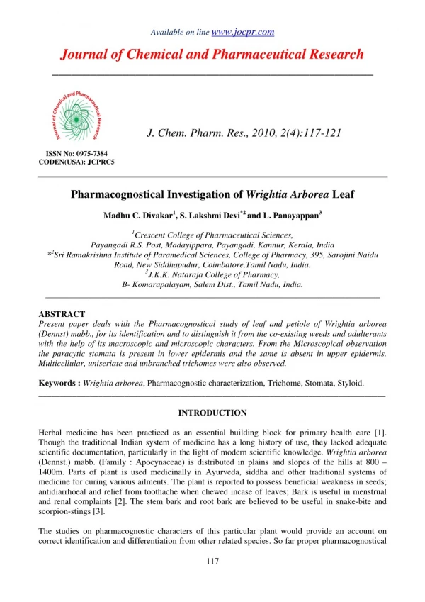
Present paper deals with the Pharmacognostical study of leaf and petiole of Wrightia arborea<br>(Dennst) mabb., for its identification and to distinguish it from the co-existing weeds and adulterants<br>with the help of its macroscopic and microscopic characters. From the Microscopical observation<br>the paracytic stomata is present in lower epidermis and the same is absent in upper epidermis.<br>Multicellular, uniseriate and unbranched trichomes were also observed.

E N D
Available on line www.jocpr.com Journal of Chemical and Pharmaceutical Research __________________________________________________ J. Chem. Pharm. Res., 2010, 2(4):117-121 ISSN No: 0975-7384 CODEN(USA): JCPRC5 Pharmacognostical Investigation of Wrightia Arborea Leaf Madhu C. Divakar1, S. Lakshmi Devi*2 and L. Panayappan3 1Crescent College of Pharmaceutical Sciences, Payangadi R.S. Post, Madayippara, Payangadi, Kannur, Kerala, India *2Sri Ramakrishna Institute of Paramedical Sciences, College of Pharmacy, 395, Sarojini Naidu Road, New Siddhapudur, Coimbatore,Tamil Nadu, India. 3J.K.K. Nataraja College of Pharmacy, B- Komarapalayam, Salem Dist., Tamil Nadu, India. ______________________________________________________________________________ ABSTRACT Present paper deals with the Pharmacognostical study of leaf and petiole of Wrightia arborea (Dennst) mabb., for its identification and to distinguish it from the co-existing weeds and adulterants with the help of its macroscopic and microscopic characters. From the Microscopical observation the paracytic stomata is present in lower epidermis and the same is absent in upper epidermis. Multicellular, uniseriate and unbranched trichomes were also observed. Keywords : Wrightia arborea, Pharmacognostic characterization, Trichome, Stomata, Styloid. _________________________________________________________________________________ INTRODUCTION Herbal medicine has been practiced as an essential building block for primary health care [1]. Though the traditional Indian system of medicine has a long history of use, they lacked adequate scientific documentation, particularly in the light of modern scientific knowledge. Wrightia arborea (Dennst.) mabb. (Family : Apocynaceae) is distributed in plains and slopes of the hills at 800 – 1400m. Parts of plant is used medicinally in Ayurveda, siddha and other traditional systems of medicine for curing various ailments. The plant is reported to possess beneficial weakness in seeds; antidiarrhoeal and relief from toothache when chewed incase of leaves; Bark is useful in menstrual and renal complaints [2]. The stem bark and root bark are believed to be useful in snake-bite and scorpion-stings [3]. The studies on pharmacognostic characters of this particular plant would provide an account on correct identification and differentiation from other related species. So far proper pharmacognostical 117
S. Lakshmi Devi et al J. Chem. Pharm. Res., 2010, 2(4): 117-121 ________________________________________________________________________________ studies for leaves of Wirghtia arborea (Dennst) mabb. have not been reported and hence our efforts were devoted in this detection. EXPERIMENTAL SECTION Materials and Methods The plant was collected from shevaroy hills, Tamil Nadu and identified and authenticated (BSI/SC/5/23/08-09/Tech) by Botanical Survey of India, Tamil Nadu Agricultural University, Coimbatore. And the specimen was deposited in the herbarium. Anatomical studies were carried out as per standard methods [4]. For anatomical studies, sections of about 10-12µm thickness were prepared and stained with polychromatic stain, toluidine blue and stained with safranin and fast green[5]. Stomatal index was calculated as per standard methods[6,7]. Micro photographs at different magnifications were taken with Nikon Lab photo 2 microscopic unit to study the anatomical characters. Polarized light was employed to study the nature of crystals and styloids [8]. RESULTS AND DISCUSSION The macroscopical studies revealed the shape of leaves as elliptic-oblong with entire margin, acute base 6mm long petiole, slightly bitter in taste and size varying form 6cm wide and 15cm long. In the microscopical studies, the plant showed the presence of paracytic type of stomata (Fig -1), multicellular, uniseriate and unbranched trichomes (Fig – 2a, 2b). Fig. I Paradermal section of the epidermis {Abaxial epidermis} Legend for the figure (BC - Basal Cell of the epidermal trichome; RC - Rosette cells; SC – subsidiary cells; St – stoma) Fig IIa, IIb : Powder Microscopy 2a. One trichome magnified Legend for the figures (BC – Basal cell; CL – Cell Lumen; CW – Cell wall; TP – Terminal part ) 2b. A trichome with its basal cell 118
S. Lakshmi Devi et al J. Chem. Pharm. Res., 2010, 2(4): 117-121 ________________________________________________________________________________ The lamina is bifacial with distinct palisade spongy mesophyll. The palisade zone has two layers of cylindrical wide cells, this is 50µm thick, 3 or 4 layered spongy parenchyma. Mid-rib region showed the ground tissue 2-3 layers of collenchyma and few inner layers of parenchyma (Fig – 3a, 3b). Single collateral vascular strand with wide zone of parenchymatous bundle sheath. Fig.3a, 3b 3a. T.S of leaf through the midrib Legend for the figures (AbS - Abaxial Side; AdS - Adaxial Side; Abe- Abaxial Epidermis; Ade -Adaxial Epidermis; La - Lamina ; Lv- Lateral Vein; PM- Palisade Mesophyll; SM – Spongy Mesophyll; St- Styloid; MR - Midrib ; VB – Vascular Bundle) The petiole showed dispersed scattered laticifers among the ground tissue. The vascular strand of the petiole has wide bowl shaped strand. Angular xylem occurs in fairly long parallel close lines and phloem masses occur both on the inner and outer portions of the xylem which is 20µm wide (Fig -4). Fig.. IV Anatomy of the Petiole TS of petiole – entire view Legend for the figure (AB: Accessory Bundles ; AbS: Abaxial Side ; AdS - Adaxial Side; GT Ground Tissue; IPh: Inner Phloem; Lf : Laticifers; MB: Main Bundle; OPh: Outer Phloem; X: Xylem ) The powder microscopy revealed the presence of styloid and prismatic crystals (Fig- 5a, 5b), paracytic stomata, wide bowl shaped vascular strand, angular xylem and masses of phloem, slightly thick walled circular laticifers, uniseriate multicellular and unbranched trichomes. Fig. III Fig. III b 3b. T.S of lamina through lateral vein 119
S. Lakshmi Devi et al J. Chem. Pharm. Res., 2010, 2(4): 117-121 ________________________________________________________________________________ Fig. 5b Fig. V a Fig.5a, 5b. Crystals in the leaf 5a – Styloids lying along the veins 5b – Crystals in the ground parenchyma of the leaf. Legend for the figures (Cr – Crystals, MT – Mesophyll Tissue, ST – Styloids, Ve- Veins) Quantitative microscopy revealed the presence of stomata only in lower epidermis of Wrightia arborea (Dennst) mabb and the stomatal values are given in (Table -1). Table – 1: Quantitative Microscopy Of Leaves Of Wrightia Arborea PARTICULARS STOMATAL NUMBER Upper epidermis Lower epidermis STOMATAL INDEX Upper epidermis Lower epidermis (-) Represents the absence of stomata. The pharmacognostical characters reported in this work can serve as a valuable source of information and provide suitable diagnostic tool for the standardization as well as adulterant identification of this medicinal plant in future investigation or application. It will also be immense using carrying out further research and revalidation of its use. The microscopic features could help in laying down micro morphological standards as per WHO guidelines for authentication of the original drug. Acknowledgement We greatly acknowledge Dr. T.K. Ravi, Principal, College of Pharmacy, Sri Ramakrishna Institute of Paramedical Sciences, Coimbatore for the valuable support and encouragement throughout the study. WRIGHTIA ARBOREA - 220 - 23.15 120
S. Lakshmi Devi et al J. Chem. Pharm. Res., 2010, 2(4): 117-121 ________________________________________________________________________________ REFERENCES [1]OA Ondyade ; JJC Scheffer; AB Svendsen. Planta Medica., 1990, 56, 503 – 504. [2]A Chatterjee; SC Pakrashi, The Treatise of India Medicinal Plant, Vol. 4, National Institute of Science Communication and Information Resources, New Delhi, 2003; 125-127. [3]KP Kiritikar.; BD Basu. Indian Medicinal Plants, Vol. 3, International Book Distributors, Dehradun, 1981;611-612. [4]DA Johansen. Plant Microtechnique, McGraw – Hill Book Co, New York 1940; 523. [5]TP O'Brien; N Feder ; ME Mcull. Polychromatic Staining of Plant Cell Walls by Toluidine Blue – O, Protoplasma, 59,1964; 364-373. [6]TE Wallis. Text Book of Pharmacognosy, CBS Publishers and Distributors, Shahdara , New Delhi, India, 1985 ;652. [7]CK Kokate. Practical Pharmacognosy, 4th Edition, Vallabh Prakashan, Delhi, 1994;115 -123. [8]JE Sass . Elements of Botanical Microtechnique, McGraw – Hill Book Co., Inc, New York, 1940;.222. 121