Download
1 / 5
70 likes | 190 Views
Limestone is made of calcium carbonate. An Attempt is made to convert calcium carbonate to calcium oxide for<br>precursor of calcium in hydroxyapatite synthesis. Phosphate was from hydrogen phosphate [H3PO4]. Diammonium<br>hydrogen phosphate [(NH4)2HPO4] were used as precursors and ammonia was used as the agent for pH<br>adjustment. The synthesized samples was characterized by Fourier Transform Infra Red (FTIR), X-ray Difraction<br>(XRD), Scanning Electron Microscopy (SEM) and Energy Dispersive X-ray Spectroscopy (EDS). Result show that<br>hydroxyapatite with calcium oxide 1 M was give fine produce. Analysis of BET, result show Intercept of 1.174e 01<br>and slope of 250.030, total pore volume obtained from hydroxyapatite 3.881e-02 cc/g and average pore radius of<br>5.83490e 01 Ã…. <br>

E N D
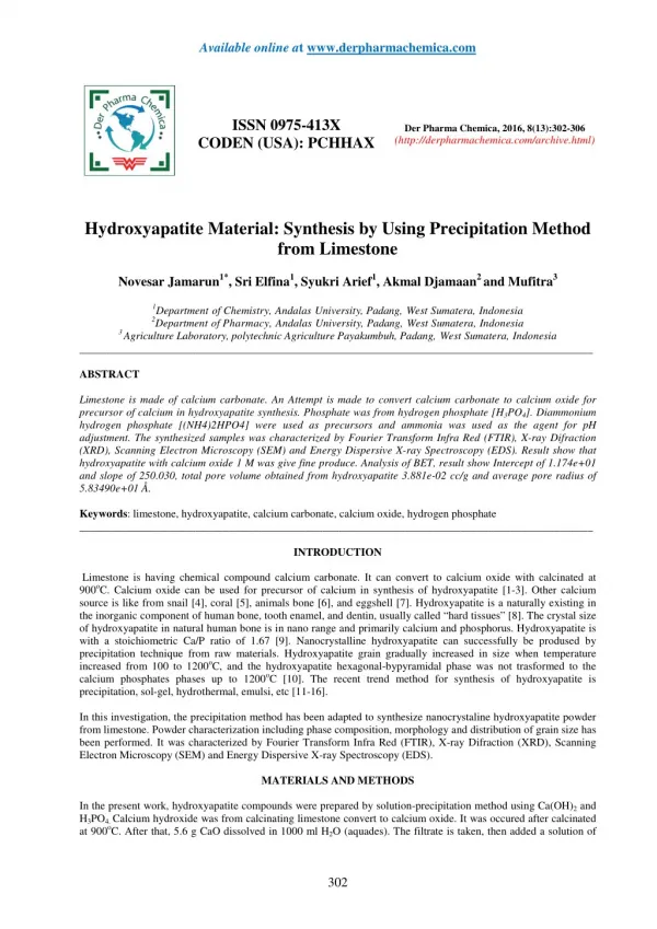
Available online at www.derpharmachemica.com ISSN 0975-413X CODEN (USA): PCHHAX Der Pharma Chemica, 2016, 8(13):302-306 (http://derpharmachemica.com/archive.html) Hydroxyapatite Material: Synthesis by Using Precipitation Method from Limestone Novesar Jamarun1*, Sri Elfina1, Syukri Arief1, Akmal Djamaan2 and Mufitra3 1Department of Chemistry, Andalas University, Padang, West Sumatera, Indonesia 2Department of Pharmacy, Andalas University, Padang, West Sumatera, Indonesia 3 Agriculture Laboratory, polytechnic Agriculture Payakumbuh, Padang, West Sumatera, Indonesia _____________________________________________________________________________________________ ABSTRACT Limestone is made of calcium carbonate. An Attempt is made to convert calcium carbonate to calcium oxide for precursor of calcium in hydroxyapatite synthesis. Phosphate was from hydrogen phosphate [H3PO4]. Diammonium hydrogen phosphate [(NH4)2HPO4] were used as precursors and ammonia was used as the agent for pH adjustment. The synthesized samples was characterized by Fourier Transform Infra Red (FTIR), X-ray Difraction (XRD), Scanning Electron Microscopy (SEM) and Energy Dispersive X-ray Spectroscopy (EDS). Result show that hydroxyapatite with calcium oxide 1 M was give fine produce. Analysis of BET, result show Intercept of 1.174e+01 and slope of 250.030, total pore volume obtained from hydroxyapatite 3.881e-02 cc/g and average pore radius of 5.83490e+01 Å. Keywords: limestone, hydroxyapatite, calcium carbonate, calcium oxide, hydrogen phosphate _____________________________________________________________________________________________ INTRODUCTION Limestone is having chemical compound calcium carbonate. It can convert to calcium oxide with calcinated at 900oC. Calcium oxide can be used for precursor of calcium in synthesis of hydroxyapatite [1-3]. Other calcium source is like from snail [4], coral [5], animals bone [6], and eggshell [7]. Hydroxyapatite is a naturally existing in the inorganic component of human bone, tooth enamel, and dentin, usually called “hard tissues” [8]. The crystal size of hydroxyapatite in natural human bone is in nano range and primarily calcium and phosphorus. Hydroxyapatite is with a stoichiometric Ca/P ratio of 1.67 [9]. Nanocrystalline hydroxyapatite can successfully be prodused by precipitation technique from raw materials. Hydroxyapatite grain gradually increased in size when temperature increased from 100 to 1200oC, and the hydroxyapatite hexagonal-bypyramidal phase was not trasformed to the calcium phosphates phases up to 1200oC [10]. The recent trend method for synthesis of hydroxyapatite is precipitation, sol-gel, hydrothermal, emulsi, etc [11-16]. In this investigation, the precipitation method has been adapted to synthesize nanocrystaline hydroxyapatite powder from limestone. Powder characterization including phase composition, morphology and distribution of grain size has been performed. It was characterized by Fourier Transform Infra Red (FTIR), X-ray Difraction (XRD), Scanning Electron Microscopy (SEM) and Energy Dispersive X-ray Spectroscopy (EDS). MATERIALS AND METHODS In the present work, hydroxyapatite compounds were prepared by solution-precipitation method using Ca(OH)2 and H3PO4. Calcium hydroxide was from calcinating limestone convert to calcium oxide. It was occured after calcinated at 900oC. After that, 5.6 g CaO dissolved in 1000 ml H2O (aquades). The filtrate is taken, then added a solution of 302
Novesar Jamarun et al _____________________________________________________________________________ Der Pharma Chemica, 2016, 8 (13):302-306 100 ml 0.06 M hydrogen phosphated, H3PO4, slowly while stirring for 1 hour at 25°C (room temperature) at pH, 11. The addition of ammonium hydroxide (NH4OH) to pH adjustment. 10Ca(OH)2 + 6 H3PO4? Ca10(PO4)6(OH)2 + 18H2O The solution formed was precipitated for 1 day or +15 hours until a precipitate is formed. The precipitate that formed was filtered and dried at a temperature of + 110oC aiming to eliminate solvents that still contained. The precipitate which has been dried and then ground into powder, then calcinated at 900oC for 2 hours. Nanopowder formed then characterized with FTIR, XRD and SEM. RESULTS AND DISCUSSION analysis of FTIR Infrared characteristic was carried out on two samples to study the spectra characteristics indivicates of the chemical bonding in the synthesized hyroxyapatite. The spectrum can be dividedinto some regions with peaks having wave numbers around 3437, 3448, and 3571 cm-1 are due prensence of –OH bond. The peaks observed around at 1042, 1034, 1089 and 962 cm-1 are due to the precence of P-O bonds in phosphate groups. Thus, the presence of PO4 group in hydroxyapatite is almost confirmed from IR studies [1,2,15]. Analysis of XRD Fig.2 shows the XRD pattern of hyroxyapatite 0.1 M and 1 M. It can be seen from the results of XRD two patterns was different (Fig.2). Hyroxyapatite 0,1 M suitability with hydroxyapatite ICSD 26205 standart (Fig.-3). The presence of noise contained in the spectrum indicates the presence of amorphous hydroxyapatite. while the sharp spectrum indicates the formation of hydroxyapatite crystals. Analysis of SEM-EDX SEM observation was performed at State University of Malang (UNM). The images was observed to be almost like spherical in agglomerate particles. The uniform grain size with narrow size distribution corresponding to the cristallinity improvement of HA 0.1 M powders. The result of measurement of elemental composition (Ca and P content) and Ca/P molar ratio are summarized in Table 1. The Ca/P stoichiometry of 0.1 M was analysed by EDX. It shows that HA powder with Ca/P ratio is below ratio 1.67, indicate other compound in sample [2,7]. Analysis of BET Analysis of the surface area of the hydroxyapatite synthesized from limestone, carried out using equipment Surface Area Analyzer (SAA). Obtained hydroxyapatite surface area of 13.304. From the analysis of BET, to use nitrogen gas adsorption isotherms, adsorption isotherms obtained chart below. Table 1. Ca and P content in th synthesized hydroxyapatite powder and Ca/P ratio Measured content (Wt%) PK 22.90 CaK 44.65 3- Ca/P ratio Element 1.51 303
Novesar Jamarun et al _____________________________________________________________________________ Der Pharma Chemica, 2016, 8 (13):302-306 Fig. 1 FTIR spectrum of hyroxyapatite powders at (a) 0.1 M and (b) 1M Intensity (a.u) (b) (a) 10 20 30 40 50 60 0) 2 θ( Fig. 2. XRD patterns of HA powders at (a) 0.1 M and (b) 1M 304
Novesar Jamarun et al _____________________________________________________________________________ Der Pharma Chemica, 2016, 8 (13):302-306 standart 26205 Intensity (a.u.) HA 0.1 M 10 20 30 40 50 60 70 80 2 θ(0) Fig. 3. XRD pattern of HA powders synthesized with HA standart ICDS 26205 Fig. 4. SEM images of HA powder at 0,1 M Fig. 5. Isoterm Adsorption-Desorption nitrogen gas of the hidroxyapatite Intercept of 1.174e+01 and slope of 250.030, total pore volume obtained from hydroxyapatite 3.881e-02 cc/g and average pore radius of 5.83490e+0 305
Novesar Jamarun et al _____________________________________________________________________________ Der Pharma Chemica, 2016, 8 (13):302-306 CONCLUSION Precipitation method was used in the present hydroxyapatite to its simplicity as well as aconomical benefit it offers on industrial scale. The precursor calcium from calcinating limestone convert to calcium oxide dissolved H2O (aquades) can result hydroxyapatite. Hyroxyapatite 0,1 M was better than hydroxyapatite 1 M. It Ca/P ratio is below ratio 1.67, indicate other compound in sample Analysis of the surface area from BET, Intercept of 1.174e+01 and slope of 250.030, total pore volume obtained from hydroxyapatite 3.881e-02 cc/g and average pore radius of 5.83490e+01 Å. Acknowledgements The authors thank Andalas University, Republic of Indonesia, because several parts of this work are supported by LPPM andalas University, that is under HKRGB, and Research Grant No.46//UN.16/HKRGB/LPPM/2016. REFERENCES [1]T.P. Sari, N Jamarun, Syukri, Z. Azharman , A. Asregi, Orient. J. Chem., 2014, 30, 1799-1804. [2]N. Jamarun, A. Zefri, A. Syukri, T.P. Sari, A. Asregi, Elfina S, Rasayan J. Chem., 2015, 8 (1). 133-137 [3]N. Jamarun, R. Juita and J. Rahayuningsih, Research Journal of Pharmaceutical, Biological and Chemical Sciences., 2015, 6(5), 136-140. [4]M.D. Adak, K.M. Purohit, Trends Biomater. Artif. Organs, 2011, 25, 101-106, [5]S. Zamani, E. Salahi, I. Mobasherpour, Canadian Chemical Transactions, 2013, 1, 173-190, [6]S. Sobczak, Zygmunt Kowalski, and Zbigniew Wzorek.; Act. Bioeng. & Biomech., 2009, 11, 23-28 [7]Y. Aziz, N. Jamarun, S. Arief and H.Nur, Orient. J. Chem., 2015, 31, 1099-1105. [8]H. Eslami, M. Solati-hashjin, M. Tahriri, Iranian Journal of Pharmaceutical Sciences, 2008, 4, 127-134. [9]S.S. Abidi, Q. Murtaza, Journal of Materials Science & Technology, 2014, 30, 307-310. [10]D. Meza, I. Figueroa, C. Flores-Morales, & C, P.-B. M.. Revista Mexicana de Fısica, 2011, 57, 471-474. [11]A.K. Nayak, International Journal of Chemtech Research, 2010, 2, 903-907. [12]O.R. Bingöl and C. Durucan, Americ. J. Bio. Sci., 2012, 4, 50-59 [13]N. Jamarun, T.P. Sari, S. Arief, Z. Azharman, A. Asril, Research Journal of Pharmaceutical, Biological and Chemical Sciences, 2015, 6 (3).1065-1069 [14]Dalong Li, Xin Huang, Yadong Wu, Jiwei Li, Weilu Cheng, Jinmei He, HuayuTian and Yudong Huang, Biomater. Sci., 2016, 4, 272-280. [15]N. Jamarun, A. Asril, Zulhadjri, Z. Azharman, and T.P. Sari., J. Chem. Pharm. Res., 2015, 6 (4). 832-837 [16]M. Xin, H. Zhiwei, H. Fengxuan, Z. Zhiyuan, C. Liang, L. Bin,Colloids and Surfaces B: Biointerfaces. 2016, 143, 81-87 306