Download
1 / 12
120 likes | 159 Views
Doxorubicin (Adriamycin) is a potent chemotherapeutic agent for the treatment of a variety of human malignancies. Among several different mechanisms proposed for explaining the<br>pathogenesis, increasing oxidative stress appears to play a major model that has been shown to increase free radical producing and decrease activities of endogenous antioxidant enzymes.There is an early depression in glutathione enzymes such as reduced glutathione, glutathioneperoxidase and glutathione-s-transferase. Cardiomyopathy is also associated with apoptosis which precede the development of cardiotoxicity and congestive heart failure. Many different approaches have been used to minimize the incidence of doxorubicin-induced cardiomyopathy. In the present study, the rabbit model, the development of cardiomyopathy was prevented by<br>reducing oxidative stress using natural antioxidant vitamin E (50 IU/kg body weight) and flavonoids morin, rutin and quercetin, which are generally available in the natural diet.

E N D
Available on line www.jocpr.com Journal of Chemical and Pharmaceutical Research __________________________________________________ J. Chem. Pharm. Res., 2010, 2(3):754-765 ISSN No: 0975-7384 CODEN(USA): JCPRC5 Cardioprotective effects of vitamin E, morin, rutin and quercetin against Doxorubicin induced oxidative stress of rabbits: A biochemical study 1*Raja Kumar Parabathina, 2G. Vijaya Raja, 3M. Nageswara Rao, 4G. Srinivasa Rao and 5Kaza Somasekhara Rao 1Department of Biochemistry, Acharya Nagarjuna University, Guntur, Andhra Pradesh, India 2Department of Biotechnology, Acharya Nagarjuna University, Guntur, Andhra Pradesh, India 3Department of Chemistry, Sri Chaitanya Mahila Kalasala, Vijayawada, Andhra Pradesh, India 4Department of Pharmacology and Toxicology, NTR College of Veterinary Science, Gannavaram, Andhra Pradesh, India 5Department of Chemistry, ANU-PG Campus, Nuzvid, Andhra Pradesh, India ______________________________________________________________________________ ABSTRACT Doxorubicin (Adriamycin) is a potent chemotherapeutic agent for the treatment of a variety of human malignancies. Among several different mechanisms proposed for explaining the pathogenesis, increasing oxidative stress appears to play a major model that has been shown to increase free radical producing and decrease activities of endogenous antioxidant enzymes. There is an early depression in glutathione enzymes such as reduced glutathione, glutathione peroxidase and glutathione-s-transferase. Cardiomyopathy is also associated with apoptosis which precede the development of cardiotoxicity and congestive heart failure. Many different approaches have been used to minimize the incidence of doxorubicin-induced cardiomyopathy. In the present study, the rabbit model, the development of cardiomyopathy was prevented by reducing oxidative stress using natural antioxidant vitamin E (50 IU/kg body weight) and flavonoids morin, rutin and quercetin, which are generally available in the natural diet. The four weeks treatment of flavonoids (20mg/kg body weight) were affectively ameliorated the oxidative stress induced cardiomyopathy in rabbits by doxorubicin (10mg/kg body weight) treatment for two days. The flavonoids alter the biochemical parameters at optimum levels. The oxidative markers (lipid peroxidation, catalase, reduced glutathione, and glutathione-s-transferase) were significantly maintained by the flavonoids in blood. The overall conclusion of this study is that the flavonoids can act as antioxidants in the body and they can protect the biochemical and oxidative markers at optimum levels. By this study, the author suggests that rabbits are good experimental models to conduct various types of experiment in Biochemistry and Pharmacology, 754
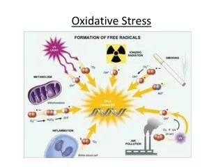
Raja Kumar Parabathina et al J. Chem. Pharm. Res., 2010, 2(3):754-765 _____________________________________________________________________________ and the diet rich flavonoids can useful in the protection of certain diseases like cardiomyopathy (CAD, hypertension, heart failure and stroke), diabetes, cancers and oxidative stress etc., Key words: Oxidative stress, cardiomyopathy, doxorubicin, morin, rutin, quercetin. ______________________________________________________________________________ INTRODUCTION Cardiovascular diseases (coronary artery disease, hypertension, heart failure, and stroke) are the leading causes of death in human beings of modern days. Oxidative stress is the unifying mechanism for many cardiovascular risk factors (diabetes and obesity) [1]. Population studies have shown that up to 80% of cardiovascular diseases, 90% of type II diabetes and approximately 30% of cancers are due to generation of free radicals under pathogenic conditions that could be avoided by diet and life style changes [2, 3]. The organ systems most susceptible to oxidative damage are the pulmonary system, the brain, the eye, the circulatory and the reproductive system. Oxidative damage causes a net stress on the normal body functions and may result in developing of many specific diseases [4]. Many free radicals exist in living systems are unstable and highly reactive, and disrupt the equilibrium of biological systems by damaging their major constituent molecules, leading eventually to cell death [4-7] These radicals can be released from NADP oxidase, xanthine oxidase, lipoxygenase, mitochondria, or the uncoupling of nitric oxide synthase in vascular cells and damage cell lipids, membranes, proteins and DNA (Leila Bettina Seres, 2006). Free radicals can also react with serum LDL and resultant oxidized LDL damages the arterial wall [4]. To compensate for this, the cells have evolved both the enzymatic and non-enzymatic mechanisms to protect against oxidant’s toxic effects. The dose related cardiomyopathy and congestive heart failure due to doxorubicin has limited the use of this drug [8, 9]. The production of superoxide radical ions by oxidation-reduction cycling is critical in mediating the chronic cardio toxicity associated with the clinical use of DOX. Redox cycling of doxorubicin-derived quinine-semiquinone, in the presence of oxygen, and transition metals, yields superoxide radicals. Increased levels of oxygen species due to doxorubicin have been observed directly by electron spin resonance spectroscopy [10, 11] and indirectly by an increase in tissue malondialdehyde (MDA) which is a breakdown product of lipid peroxidation [12-14]. During doxorubicin treatment serum enzymes such as SGOT, LDH, and CPK were increased significantly, particularly in the late stages of cardiac failure [8, 15-18] mice [19, 20], and rats [21, 22]. It is well documented that the major side effect, cardiomyopathy during doxorubicin treatment in malignancies is due to oxidative stress caused by release of free radicals. In heart GSHPx is extremely important because of its ability to use and remove organic and inorganic peroxides [13]. Administration of doxorubicin resulted in a dose and time dependent decrease in myocardial glutathione enzymes activity in rabbits and mice [23-27]. Adjunct therapy of doxorubicin along with antioxidants has become a practice to curb the oxidative damage by doxorubicin in recent times. Flavonoids are polyphenolic compounds present in the many plant derived foods. Many epidemiological studies have shown that flavonoid intake is inversely related to mortality from coronary heart disease and to the incidence of heart attacks. In a recent study conducted in the Netherlands suggested that flavonoids can replace vitamin E as chain-breaking antioxidants in 755
Raja Kumar Parabathina et al J. Chem. Pharm. Res., 2010, 2(3):754-765 _____________________________________________________________________________ liver microsomal membranes. The capacity of flavonoids to act as antioxidants depends upon their molecular structure. The position of hydroxyl groups and other features in the chemical structure of flavonoids are important for their antioxidant and free radical scavenging activities. Morin, rutin and quercetin by acting as antioxidants exhibited several beneficial effects, such as anti-inflammatory, antiallergic, antiviral as well as an anticancer activity. They have also been suggested to play a protective role in liver diseases, cataracts, and cardiovascular diseases. Quercetin acting as free radical scavengers where shown to exert a protective effect in reperfusion ischemic tissue damage [28-34]. According to their specificity in antioxidation function, certain selected flavonoids naturally available in diet (morin, rutin and quercetin) are used in the present study to investigate their ameliorative affects on biochemical studies doxorubicin induced oxidative stress cardiomyopathy in rabbits. Keeping in view the present study was aimed at exploration of amelioration effect of different flavonoids (morin, rutin, and quercetin) in doxorubicin induced cardiomyopathy oxidative stress. The aim and objective of the present study is to study biochemical changes in doxorubicin induced cardiomyopathy in rabbits. To study the effects of flavonoids (morin, rutin, and quercetin) on biochemical in cardiomyopathy induced by the doxorubicin. EXPERIMENTAL SECTION Chemicals and reagent kits The chemicals used in the present study were of analytical grade from E.Merck (India), SISCO Laboratories and Loba Chemicals and some chemicals were procured from Sd Fine Chemicals, Navi Mumbai, India. Vitamin E (Bio-E 400) was procured from the Dr.Reddy’s Laboratories, Hyderabad and drug Doxorubicin (Doxopar-50) Parenteral drugs (India) limited, Indore, India. Some of the reagent kits were purchased from by the Laboratory of Ensure Biotech Pvt Limited, Hyderabad, and few reagent kits were purchased from the Transonic Bio-Medicals Limited, Solan (HP), was used for this study. Animals: Thirty apparently healthy, New Zealand white rabbits weighing 2.5 to 3.0 kg (about 3-6 months) were obtained from Laboratory of small animal house, Department of Pharmacology, Dr.Pinnamaneni Siddhartha Institute of Medical Sciences and Research Foundation, Chinnoutapalli, Gannavaram, Krishna District, Andhra Pradesh, India. The animals were housed in the cages of departmental laboratory animal shed. All the animals were fed with control diet during a month acclimatization period. All animals were kept under uniform managerial and standard hygienic conditions through the experimental period. All the rabbits were weighed and randomly housed, two animals in each cage. The cages were located in a well ventilated house and all the animals had free access to feed and water at all times. Experimental Design: Thirty rabbits were randomly divided into five groups and six animals in each group. The group I animals are Controls which were fed normal diet about twenty eight days and the twenty ninth and thirtieth day i.e. two days doxorubicin 10 mg /kg body weight was given intra venously, for the group II animals antioxidant vitamin E 50 IU/ kg body weight was given orally along with normal diet for twenty eight days and the two doses of doxorubicin was administrated on twenty ninth and thirtieth day, for the group III, IV & V were fed orally with flavonoids morin, rutin and quercetin, 20mg/ kg body weight along with ad libitum of water and diet respectively for twenty 756
Raja Kumar Parabathina et al J. Chem. Pharm. Res., 2010, 2(3):754-765 _____________________________________________________________________________ eight days and the doxorubicin was administrated in two doses on twenty ninth and thirtieth day as in group I and II rabbits. At weekly intervals blood samples were collected and analyzed foe hematological and biochemical (general and oxidative markers) parameters. On 5th time i.e. after doxorubicin treatment for two days, again blood samples were collected and analyzed for both parameters, then 50% of rabbits from each group were sacrificed for oxidative parameters in tissues and to study the histopathological changes in the tissues (Heart, liver and kidney). Methods: The biochemical parameters such as protein profile such as total protein(Biuret method), albumin(BCG method), globulin, A:G Ratio), lipid profile: total cholesterol (Cholesterol esterase-enzymatic method), triglycerides(Glycerol-3-phosphate oxidase- enzymatic method), HDL-cholesterol (Phosphotungstic acid cholesterol(Friedwalds formula), blood sugar (GOD-POD method), urea (Berthelot method), blood urea nitrogen, uric acid (Uricase-enzymatic method), Creatinine (alkaline picrate method), enzymes SGOT and SGPT (IFCC method), ALP (p-nitrophenyl phosphate method), minerals sodium (Modified Maruna and Trinders method), potassium (Turbidometric method), phosphorus (Fiske and Subbarow method) and oxidative parameters Catalase (Bergmeyer method), reduced glutathione (DTNB method), glutathione-s-transferase (habig method) and lipid peroxidation (Stock and Dormandy method) were estimated with standard protocols. RESULTS AND DISCUSSION From the table - 1 it is evident that vit E increased total protein and albumin levels, in remaining all group total protein, albumin and globulin levels were decreased except in case of quercetin in which albumin and globulin were increased. According this study, the globulin levels were more compared with the albumin concentration, indicates that the rabbits had more immunopotency rather than the muscle formation. TABLE: 1. Concentration of protein profile in the groups of rabbits before and after the treatment of doxorubicin (the before values are average values of 4th week treatment of vitamin-E and flavonoids morin, rutin and quercetin) method), LDL-cholesterol and VLDL- Total Protein Before 9.15±0.37 9.56±0.33 9.03±0.33 9.36±0.51 Albumin Globulin After 8.8±0.47 10.35±0.61 8.98±0.15 9.01±0.32 7.84±0.42 Before 2.58±0.15 2.38±0.15 2.61±0.20 2.48±0.16 3.75±0.30** 3.93±0.36** 3.33±0.34** After 2.56±0.10 2.55±0.12 2.51±0.17 2.43±0.15 Before 6.56±0.25 7.18±0.43 6.41±0.37 6.88±0.57 After 6.23±0.52 7.8±0.63 6.46±0.24 6.58±0.46 3.91±0.28** Control Vit-E Morin Rutin Quercetin 7.08±0.56** * In a row differ significantly at P<0.05, ** In a row differ significantly at P< 0.01 The lipid profile included in the study was total Cholesterol, Triglycerides, HDL-Cholesterol, LDL-Cholesterol and VLDL-Cholesterol was shown various changes on the treatment of DOX and flavonoids in this study. Table- 2 shows that morin and quercetin decreased total cholesterol where as increased levels were found in rutin, vit E and control group. Morin, rutin, vit E increased triglycerides and decreased in quercetin group but levels were maintained normal in DOX treated control group. Morin and quercetin decreased LDL levels where as vit E increased but no significant changes found in rutin and control group animals. Quercetin and control group animals showed decreased HDL levels and there is no significant change was found in Morin, rutin and vit E animals. VLDL levels were increased in morin, rutin and Vit E group but decreased in quercetin and maintained normal in control group. The overall study on lipid profile 757
Raja Kumar Parabathina et al J. Chem. Pharm. Res., 2010, 2(3):754-765 _____________________________________________________________________________ concentration indicates that pretreatment of flavonoids was able to reduce the cardiac toxicity by reducing the lipid profiles in the rabbits even after the treatment of DOX. Pre treatment of vitamin E, flavonoids showed the reduction in lipid profile with concomitantly increase Fig: 1. Concentration of protein profile in the groups of rabbits before and after the treatment of doxorubicin (the before values are average values of 4th week treatment of vitamin-E and flavonoids morin, rutin and quercetin) TABLE: 2. Concentration of lipid profile in the groups of rabbits before and after the treatment of doxorubicin (the before values are average values of 4th week treatment of vitamin-E and flavonoids morin, rutin and quercetin) Total Cholesterol Triglycerides Before After Before After Before HDL LDL VLDL After Before After Before After Control 119.69± 1.76 121.48± 2.78 115.59± 2.47 115.16± 3.45 31.02± 0.49 28.53± 0.59 65.55± 1.70 69.91± 3.02 23.11± 1.97 23.03± 2.76 90.31± 3.55** 104.04± 1.90** 94.79± 1.69** 115.95± 2.85 28.67± 0.63 29.53± 0.47 42.67± 3.88** 51.31± 2.45** 18.95± 0.33 23.19± 2.28 Vit-E Morin 136.09± 2.85** 121.66± 2.06 111.97± 1.83 147.94± 2.77** 27.23± 0.64 27.32± 0.22 86.46± 2.97* 64.75± 2.08 22.39± 1.46 29.58± 2.21 116.74± 1.84 126.45± 2.34 112.91± 2.08 154.1± 2.72** 28.25± 0.42 28.27± 0.39 65.91± 2.21 67.36± 1.84 22.58± 1.67 30.82± 2.18 Rutin Quercetin 125.20 ± 2.54** 202.24± 2.66** 164.79± 2.33** 178.81± 2.83** 164.94± 5.41** 41.27± 1.53** 38.51± 1.16 93.28± 1.70** 35.76± 2.26** 32.98± 1.08** * In a row differ significantly at P<0.05, ** In a row differ significantly at P< 0.01 758
Raja Kumar Parabathina et al J. Chem. Pharm. Res., 2010, 2(3):754-765 _____________________________________________________________________________ Fig: 2. Concentration of lipid profile in the groups of rabbits before and after the treatment of doxorubicin (the before values are average values of 4th week treatment of vitamin-E and flavonoids morin, rutin and quercetin). in HDL-cholesterol was observed. Decrease in the blood lipid profiles and increase in the HDL- Cholesterol levels in flavonoid treated groups is due to inhibition of hepatic cholesterol biosynthesis, increased fecal bile acid secretion and stimulation of receptor mediated catabolism of LDL Cholesterol and increase in the uptake of LDL-cholesterol from blood by liver [35]. In control group after the treatment of doxorubicin for 2 days at the end of 4 weeks on normal diet, reducing sugar was decreased (table-3). Vitamin E and morin treated groups were decreased the blood sugar concentration, but rutin and quercetin treated group rabbits increased the blood sugar concentration even after the treatment of doxorubicin. Depletion in the reducing sugar due to DOX may be affected the release of insulin from the pancreas, or either due to hyper metabolic consumption of energy or diseases related to liver [39]. The serum SGOT and SGPT levels were decreased in group I-V but ALP levels were increased in group IV and V and decreased in group II and III but maintained normal in control group animals (table-4). The decreased levels of SGOT and SGPT had no significant affect in the metabolism; where as increased levels may lead to severe liver disorders like necrosis and myocardial infarction, which are indicators of poor quality protein in diets fed [36]. ALP levels were also maintained in normal range that indicates no significant affect on the rabbits. Even though the values were decreased in Serum enzymes by doxorubicin treatment had no specific affects, but the flavonoids were protected at optimum levels to maintain the normal range. 759
Raja Kumar Parabathina et al J. Chem. Pharm. Res., 2010, 2(3):754-765 _____________________________________________________________________________ TABLE: 3. Concentration of blood sugar in the groups of rabbits before and after the treatment of doxorubicin (the before values are average values of 4th week treatment of vitamin-E and flavonoids morin, rutin and quercetin) Blood sugar Before 71.4±2.99 71.28±2.07 68.21±2.70 68.4±0.90 109.19±5.28** After Control Vit-E Morin Rutin Quercetin 62.11±2.23 68.48±3.22 66.35±1.80 69.78±2.39 118.74±4.97** * In a row differ significantly at P<0.05, ** In a row differ significantly at P< 0.01 Fig: 3. Concentration of blood sugar in the groups of rabbits before and after the treatment of doxorubicin (the before values are average values of 4th week treatment of vitamin-E and flavonoids morin, rutin and quercetin) TABLE: 4. Concentration of serum enzymes in the groups of rabbits before and after the treatment of doxorubicin (the before values are average values of 4th week treatment of vitamin-E and flavonoids morin, rutin and quercetin) SGOT SGPT ALP Before 102.71±2.14 106.86±2.17 77.85±2.34** 102.28±3.21 44.13±2.53** * In a row differ significantly at P<0.05, ** In a row differ significantly at P< 0.01 After 91.8±3.65 75.35±2.61** 60.43±2.06** 79.2±2.32* 30.41±3.15** Before 75.62±2.85 80.56±1.16 79.31±3.03 83.81±1.65* 41.85±1.19** After Before 67.6±1.82 72.85±2.12 83.59±1.74** 76.71±2.40* 55.36±1.06** After Control Vit-E Morin Rutin Quercetin 72.54±2.93 61.68±1.74* 66.35±2.44 55.77±2.88** 31.25±1.68** 70.21±1.35 57.56±1.37** 69.39±1.64 82.86±2.26** 64.78±1.54 760
Raja Kumar Parabathina et al J. Chem. Pharm. Res., 2010, 2(3):754-765 _____________________________________________________________________________ Fig: 4. Concentration of serum enzymes in the groups of rabbits before and after the treatment of doxorubicin (the before values are average values of 4th week treatment of vitamin-E and flavonoids morin, rutin and quercetin). Quercetin decreased the nitrogenous profile but remaining groups increased blood urea, BUN and uric acid (table-5). Increased serum urea concentration may suggest an increase in activities of urea enzymes orninthine carboxyl transferase and orginase which may also indicate kidney damage [37]. The normal range of values obtained implied therefore that the dietary proteins of the CPB diets and the control were well utilized [38]. The urea levels were low because of low intake of protein or low catabolism of muscle proteins. Generally uric acid formation was very low in terrestrial animals, but the rabbit may take less water to form uric acid in their metabolism. The nitrogenous catabolic pathways may increase the formation uric acid is due to high protein intake or nucleotide catabolism. Table-6 shows the Sodium levels were decreased in I-III and increased in IV and V. Potassium levels increased in group III, decreased in group V and maintained normal in group I, II, IV. Phosphorus levels were normal during entire experimental period in group I, III and IV and mount in group V but significantly decreased in group II. Flavonoids significantly increased sodium concentration due to increase the transport mechanism of the free radicals which were formed on DOX treatment or reduction in the water reabsorption in kidneys. The increase in potassium may be due to decrease in the reabsorption of water at kidneys or renal failure by DOX. The decreased levels of potassium in quercetin treated group may be due to malabsorption syndrome. This study indicates that the DOX-induced oxidative stress had significantly ameliorated by the flavonoids by maintaining the phosphorus concentration. Doxorubicin induced oxidative stress was confirmed in rabbits by the specific changes occurred in the biochemistry. The changes in biochemical parameters due to doxorubicin treatment in rabbits did not alter significantly. Interestingly the rabbits of group III, IV and V which were provided the flavonoids (morin, rutin and quercetin) along with diet showed remarkable antioxidant changes indicating the amelioration of oxidative stress induced by doxorubicin there by cardiomyopathy. 761
Raja Kumar Parabathina et al J. Chem. Pharm. Res., 2010, 2(3):754-765 _____________________________________________________________________________ TABLE: 5. Concentration of nitrogenous profile in the groups of rabbits before and after the treatment of doxorubicin (the before values are average values of 4th week treatment of vitamin-E and flavonoids morin, rutin and quercetin) Blood Urea Before After Before Control 12.31±0.37 13.73±0.72 5.66±0.17 Vit-E 10.86±0.72 13.75±0.52 4.99±0.33 Morin 12.25±0.59 14±0.85 5.63±0.27 Rutin 11.91±0.78 13.55±0.70 5.48±0.35 Quercetin 18.6±0.97** 14.72±0.93 8.55±0.44** * In a row differ significantly at P<0.05, ** In a row differ significantly at P< 0.01 BUN Uric acid After 6.31±0.33 6.32±0.24 6.44±0.39 6.23±0.32 6.77±0.42 Before 4.57±0.24 4.76±0.41 4.61±0.16 3.76±0.13 5.97±0.81 After 4.41±0.24 4.31±0.33 4.49±0.20 3.72±0.26 5.06±0.83 Fig: 5. Concentration of nitrogenous profile in the groups of rabbits before and after the treatment of doxorubicin (the before values are average values of 4th week treatment of vitamin-E and flavonoids morin, rutin and quercetin) TABLE: 6. Concentration of minerals in the groups of rabbits before and after the treatment of doxorubicin (the before values are average values of 4th week treatment of vitamin-E and flavonoids morin, rutin and quercetin) Sodium Potassium Phosphorus Before 6.13±0.30 6.53±0.35 6.56±0.24 6.08±0.40 5.75±0.39 Before 151.65±3.05 136.32±2.00** 116.31±1.16** 131.7±1.55** 146.31±2.04 142.78±2.38* After Before 6.56±0.64 6.72±0.21 6.07±0.46 6.87±0.56 After 6.66±0.27 6.97±0.56 8.01±0.75 6.58±0.47 3.31±0.15** After 6.02±0.16 5.91±0.31 6.27±0.38 6.03±0.25 6.36±0.25 Control Vit-E Morin Rutin Quercetin 135.03±3.95 124.45±1.99* 168.4±2.08** 176.63±3.86** 4.32±0.33** * In a row differ significantly at P<0.05, ** In a row differ significantly at P< 0.01 762
Raja Kumar Parabathina et al J. Chem. Pharm. Res., 2010, 2(3):754-765 _____________________________________________________________________________ Fig: 6. Concentration of Sodium in the groups of rabbits before and after the treatment of doxorubicin (the before values are average values of 4th week treatment of vitamin-E and flavonoids morin, rutin and quercetin) Fig: 7. Concentration of Potassium in the groups of rabbits before and after the treatment of doxorubicin (the before values are average values of 4th week treatment of vitamin-E and flavonoids morin, rutin and quercetin) Fig: 8. Concentration of Phosphorus in the groups of rabbits before and after the treatment of doxorubicin (the before values are average values of 4th week treatment of vitamin-E and flavonoids morin, rutin and quercetin) 763
Raja Kumar Parabathina et al J. Chem. Pharm. Res., 2010, 2(3):754-765 _____________________________________________________________________________ Thus, this study concludes that the flavonoids morin, rutin and quercetin can ameliorate the oxidative stress induced cardiomyopathy. Dietary intervention of flavonoids may be a good practice to protect myocardium in the doxorubicin treatment of cancers or malignancies. Flavonoids like quercetin, rutin and morin could be good adjunct molecules in doxorubicin therapy. Further investigations are necessary to prove clinical efficacy and use of flavonoids in amelioration of doxorubicin oxidative stress leading to cardiomyopathy. REFERENCES [1]Leila Bettina Seres; “Oxidative stress in cardio vascular diseases and in experimental models” PhD thesis, 2006. [2]Joint WHO/FAO Expert consultation on Diet, Nutrition and the prevention of chronic diseases draft, 2002. [3]Joye Kay Willcox, “Investigation of an Interactive Effect of Flavonoids on the Antioxidant activity of Alpha tocopherol” PhD thesis, 2002. [4]Vijayalakshmi B and Chandrasekhar M, Current trends in Biotechnology and Pharmacy., 2008, Vol (2) 239-250. [5]Ota H, Igarashi S, Hatazawa J, Janaka T, Fertil Steril., 1998, 69:303 -308. [6]Steinberg D, Parthasarathy S, Carew JE, Khoo Jc and Witztum Jc, N Engl J Med.,1989,320: 915-924. [7] Szczepanska M, Kozlik J, Skzypezak J, Miko Lajezyk M, Fertile Steril., 2003, 79: 1288- 1293. [8]Lefrak EA, Pitha J, Rosenhein S, Gottleib JA, Cancer.,1973,32: 302-314. [9]Singal PK, Iliskovic N, N Engl J Med.,1998,339: 900-905. [10]Thornally PJ, Dodd NJF, Biochem Pharma ed.,1985, 34: 669-674. [11]Iliskovic N, Hasinoff BB, Malisza KL, Li T, Danelisen I, Singal PK, Mol Cell Biochem., 1999,196: 43-49. [12]Singal PK, Deally CMR, Weinberg LE, J Mol Cell Cardiol.,1987,19: 817-828. [13]Kaul N, Siveski, Iliskovic N, Hill M, Slezak J, Singal PK, J Pharma ed Toxicol Meth.,1993, 30: 55-67. [14]Myers CE, Mc Guine WP, Liss RH, Ifrim I, Young RC, Science.,1977,197: 165-167. [15]Britshow MR, Thompron PD, Martin RP, Mason JW, bollingham ME, Harrison DC, Am J Med., 1978, 65: 823-832. [16]Jaenke RS, Fajardo LF, Am J Surg Path.,1977,1:55-60. [17]Olson HM, Young DM, Prier DJ, Le Toy AF, Reagan RL, Am J Pathol.,1974,77:439-454. [18]Jaenke RS, Lab Invest., 1974,30: 292-303. [19]Rosenhoff SH, Olson HM, Young DM, Bosssic F, Yang RC, J Nat Cancer Invest., 1975, 55: 191-194. [20]Lambertenghi, Deliliers G, Zanon PL, Pozzoli EF, Bellini O, Tumori., 1976,62: 517-528. [21]Chalscroft SCW, Gavin JB, Hender PB, Pathology.,1973,5:99-105. [22]Singal PK, Siveski-Iliskovic N, Hill M, Thomas TP, Li Y, J Mol Cell Cardiol., 1995,27: 1055-1063. [23]Revis NW, Marusic N, J Mol Cell Cardiol., 1978,10: 945-951. [24]Doroshow JH, Locker GY and Myers CE, J Clin Invest.,1980,65:128-135. [25]Ji LL, Mitchell EW, Biochem Pharmacol,1994,47: 877-885. [26]Siveski-Iliskovic N, Kaul N, Singal PK, Circulation.,1994,89:2829-2835. [27]Siveski-Iliskovic N, Hill M, Chow DA, Singal PK, Circulation., 1995,91: 10-15. [28]Santos AC, Vyemura SA, Lopes JL, Bazon JN, Mingotto FE, Cutric, Free Radic Biol Med., 1998,24: 1455-61. [29]Halliwell, B, Nutrition Reviews., 1994, 52:253-265. 764
Raja Kumar Parabathina et al J. Chem. Pharm. Res., 2010, 2(3):754-765 _____________________________________________________________________________ [30]Fraga CG, Mactino US, Ferraro GE, Coussio JF, Boveris A, Biochem Med Metabol Biol., 1987,36: 717-20. [31]Felicia VS, Najla G, Ann PC, Madeleive M, Keneeth KC, Nutr Cancer., 1996, 26: 167-81. [32]Catherire C, Male S, Esther HL, Vadimer A, Krutorslaikh, Nutr Cancer., 1996, 26: 251-63. [33]Paul P, Ritra J, Ritva S, Mackku H, Lyly T, Eero P, Arpo A, Am J Epidemiolo., 1997, 146: 223-30. [34]Fritz B, Tobias S, Albrecht K, Chaelotte B, Kent C, J Biol Chem., 1996,271: 2262-70. [35]Khanna A. Kramesh C; Kapoor N.K, Phytotherapy Res., 1996, 10: 663. [36]Facina, O.E., A.D. Ologhobo, C.O. Ayoade, G.A. Adeniran, and O.A. Adeyemi, Proc. Anim. Sci. Assoc. Nig.,1999,4: 19-22. [37]Ajagbonna, O. P., K.I. Onifed and U. Suleman, Sokoto J.Vet.Sci., 1999, 1: 36-42. [38]Reinhold, J.G., “Manual determination of total serum proteins, albumin and globulin fractions by Biuret method in: Standard methods in clinical chemistrty (Reiner M.Ed.)”, 1953, Vol. 1, Academic press, New York, pp: 88. [39]Ahamefule F.O. Eduok G.O. Usman A. Amaefule K.U. Obua B.E; Oguike S.A, Pakistan Journal of Nutrition., 2006, 5(3): 248-253. 765