Download
1 / 7
70 likes | 104 Views
5-[1-(Aryl)-5-phenyl-1H-pyrazol-3-yl]-4-phenyl-2,4-dihydro[1,2,4]triazole-3-thione derivatives<br>1a-c were reacted with chloropropanol, chloroacetone, chlorodiethyl ether and epichlorohydrine<br>to afford the corresponding acyclonucleosides 2a-c-5a-c, respectively. Also, compound 1c was<br>reacted with formaldehyde and 4-chloroaniline or piperidine to give the corresponding Mannish<br>products 6 and 7, respectively. In addition, 1c was reacted with acrylonitrile or benzoyl chloride<br>to give N-triazole derivatives 8 and 9, respectively. The structures of newly synthesized<br>compounds were confirmed by IR, 1H NMR, MS spectral data and elemental analysis. All the<br>compounds were screened for their antibacterial and antifungal activities and they showed high<br>activity compared with Ciprofloxacin and Fusidic acid as positive controls. The compounds 2b,<br>3b, 5b, 6 and 7 were screened for their anticancer activities against breast carcinoma (MCF7)<br>and cervix carcinoma (HELA) human cell lines compared with Doxorubicin positive control.<br>The detailed synthesis, spectroscopic data, antimicrobial and anticancer activities of some<br>synthesized compounds were reported.

E N D
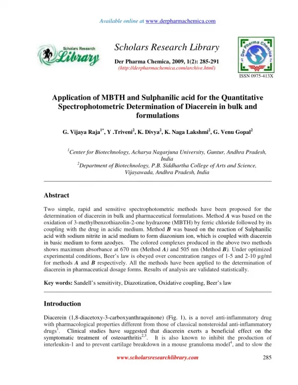
Available online at www.derpharmachemica.com Scholars Research Library Der Pharma Chemica, 2009, 1(2): 285-291 (http://derpharmachemica.com/archive.html) ISSN 0975-413X Application of MBTH and Sulphanilic acid for the Quantitative Spectrophotometric Determination of Diacerein in bulk and formulations G. Vijaya Raja1*, Y .Triveni2, K. Divya2, K. Naga Lakshmi2, G. Venu Gopal2 1Center for Biotechnology, Acharya Nagarjuna University, Guntur, Andhra Pradesh, India 2Department of Biotechnology, P.B. Siddhartha College of Arts and Science, Vijayawada, Andhra Pradesh, India ______________________________________________________________________________ Abstract Two simple, rapid and sensitive spectrophotometric methods have been proposed for the determination of diacerein in bulk and pharmaceutical formulations. Method A was based on the oxidation of 3-methylbenzothiazolin-2-one hydrazone (MBTH) by ferric chloride followed by its coupling with the drug in acidic medium. Method B was based on the reaction of Sulphanilic acid with sodium nitrite in acid medium to form diazonium ion, which is coupled with diacerein in basic medium to form azodyes. The colored complexes produced in the above two methods shows maximum absorbance at 670 nm (Method A) and 505 nm (Method B). Under optimized experimental conditions, Beer’s law is obeyed over concentration ranges of 1-5 and 2-10 µg/ml for methods A and B respectively. All the methods have been applied to the determination of diacerein in pharmaceutical dosage forms. Results of analysis are validated statistically. Key words: Sandell’s sensitivity, Diazotization, Oxidative coupling, Beer’s law ______________________________________________________________________________ Introduction Diacerein (1,8-diacetoxy-3-carboxyanthraquinone) (Fig. 1), is a novel anti-inflammatory drug with pharmacological properties different from those of classical nonsteroidal anti-inflammatory drugs1. Clinical studies have suggested that diacerein exerts a beneficial effect on the symptomatic treatment of osteoarthritis2,3. It is also known to inhibit the production of interleukin-1 and to prevent cartilage breakdown in a mouse granuloma model4, and to slow the www.scholarsresearchlibrary.com 285
G. Vijaya Raja et al Der Pharma Chemica 2009, 1 (2): 285-291 _____________________________________________________________________________ progression of cartilage lesions in a canine model of osteoarthritis5. Diacerein is known to be completely metabolized by animals and humans into rhein, an active metabolite of diacerein found in plasma and synovial fluid6. Moreover, Cruz and Pastrak have related the use of diacerein or rhein to treat and prevent vascular diseases7. Fig: 1 Chemical structure of Diacerein For its potential pharmaceutical effects on health, the development of a simple, rapid and sensitive method for the determination of diacerein in pharmaceutical preparations would therefore be highly desirable. Giannellini et al. [8] reported a validated HPLC stability-indicating method for the determination of diacerein in bulk drug substances. Han-Chun Yao et al. had proposed a Flow Injection Chemiluminescence method for the determination of diacerein at nanograms levels9. They are very few spectrophotometric methods are available till date10. Our present communication reports two simple and rapid spectrophotometric methods for the determination of diacerein in bulk and commercial dosage forms using MBTH and Sulphanilic acid as analytical reagents. The proposed reagents are used for the first time in diacerein analysis. Results and Discussion Spectral Characteristics: The method A was based on the oxidative coupling reaction of the drug with 3- methylbenzothiazolin-2-one hydrazone (MBTH) in the presence of ferric chloride in acidic medium. Method B involves diazotization of Sulphanilic acid under acidic conditions, followed by its coupling with diacerein in alkaline medium. The absorption spectra of the colored chromogens formed between Diacerein – MBTH (Method A) and Diacerein - Sulphanilic acid (Method B) have absorption maxima at 630 and 505 nm respectively. Chemistry of the colored chromogens: In the MBTH method, the drug reacts with MBTH in the presence of FeCl3 in acidic medium to give a green colored product. Actually, this is an iron catalyzed oxidative coupling reaction of MBTH with the drug. Under the reaction conditions, on oxidation, MBTH loses two electrons and one proton forming an electrophilic intermediate, which is the active coupling species. This intermediate undergoes electrophilic substitution with the drug to form the colored product. The tentative reaction scheme was given in Fig: 2 www.scholarsresearchlibrary.com 286
G. Vijaya Raja et al Der Pharma Chemica 2009, 1 (2): 285-291 _____________________________________________________________________________ In the sulphanilic acid method, diazonium salt derived from sulphanilic acid was commonly used as reagent for direct coupling procedures. The sulphanilic acid, coupling reagent was converted to their diazonium salt with HCl and sodium nitrite. Formation of diazonium salt is usually fast enough that any convenient pH between 1 and 3 can be used. The reaction is usually carried out in an ice bath, the excess nitrate is removed by reaction with ammonium sulphamate, and the pH is adjusted for the coupling reaction. The reagent is used immediately since most diazonium salts are not stable. The diazonium salt form of sulphanilic acid couples with the diacerein at alkaline pH resulting in the formation of the brown colored azodye. The tentative reaction scheme was given in the Fig: 3. Materials and Methods Experimental: Apparatus: Elico UV – Visible Double beam spectrophotometer model SL-159 with 1cm matched quartz cells was used for absorbance measurements. Materials and Reagents: All the chemicals used were of analytical grade. All the solutions were freshly prepared in distilled water. Method A: a.0.2% aqueous MBTH b.0.7% Fecl3 in 0.5% HCl. Method B: a.2% Aqueous Aniline solution b.1% Aqueous Sulphanilic acid solution c.1% Aqueous sodium Nitrite solution d. 1% Aqueous Sodium carbonate solution e.1% Aqueous Ammonium Sulphamate solution f.5N HCl Preparation of standard and sample solution: Accurately weighed 100 mg of diacerein was dissolved in 100 ml of distilled water to give a concentration of 1 mg/ml. The final concentration was brought to 100 µg/ml for both Methods A and B. General Assay procedure: Method A: Aliquots of standard drug solution containing 1-5 µg/ml of diacerein were transferred into 10ml calibrated flasks. To each flask 1.5 ml of 0.7% FeCl3 in 0.5% HCl and 1.5 ml aqueous solution of MBTH was added. The solutions were swirled and allowed to stand for 5 min. The flasks were made up to the mark with distilled water. The absorbance of the green color chromogen was measured at 670 nm against the corresponding reagent blank and calibration graph was prepared. www.scholarsresearchlibrary.com 287
G. Vijaya Raja et al Der Pharma Chemica 2009, 1 (2): 285-291 _____________________________________________________________________________ Method B: In to a series of graduated tubes add 2 ml of 1% Sulphanilic acid solution and 1 ml of 5 N Hydrochloric acid, cooled in an ice bath and 2 ml aqueous solution of 1% NaNO2 was added. The solutions were cooled to 4°C and 1 ml of 1% ammonium sulfamate was added and stirred for 5 min. 1.5 ml of 1% Na2CO3 and aliquots of standard drug solution containing 2-10 µg/ml of diacerein was added and made up to the mark with water. The solutions were mixed thoroughly and the absorbance was measured at 505 nm against reagent blank and a calibration graph was constructed. Assay of diacerein in pharmaceutical tablets: Twenty tablets were weighed and ground to a fine powder using a pestle and a mortar. The average weight of a tablet was calculated. An accurately weighed portion of the powder, equivalent to 100 mg of diacerein, was transferred into a 100 ml volumetric flask. The volume was made up to the mark with water, shaken well, and filtered through an ordinary filter paper. Convenient aliquots from this solution were taken for the determination of diacerein by the proposed methods. Analytical data: The optical characteristics such as absorption maxima, Beer’s law limits, Molar absorptivity and Sandell’s sensitivity for these methods are presented in Table-1. The regression analysis using the method of least squares was made for the slope (a) and intercept (b) obtained from different concentrations are summarized in Table-1. The precision and accuracy were found by analyzing five replicate samples containing known amounts of the drug and the results are summarized in Table-1. Fig: 2 Tentative reaction scheme for oxidative coupling of MBTH with Diacerein The accuracy of these methods was ascertained by comparing the results obtained with the proposed and reference methods in the case of formulation are presented in Table-2. As an additional check on the accuracy of these methods, recovery experiments were performed by adding known amounts of pure drug to pre-analyzed formulation and percent recovery values www.scholarsresearchlibrary.com 288
G. Vijaya Raja et al Der Pharma Chemica 2009, 1 (2): 285-291 _____________________________________________________________________________ obtained are listed in Table-3. Recovery experiments indicated the absence of interferences from the commonly encountered pharmaceutical additives and excipients. Fig: 3 Tentative reaction scheme for diazocoupling of Sulphanilic acid with Diacerein Table: 2 Optical characteristics, precision and accuracy of proposed methods MethodA Method B Parameters λmax (nm) 670 1 – 5 505 2 – 10 Beer’s law limit (µg/ ml) Sandell’s Sensitivity (µg/cm2/0.001 abs. unit) 0.0135 0.033 Molar absorptivity(Litre.mole-1.cm-1) Stability of Color (hours) Regression equation (Y)* intercept (a) slope(b) % RSD$ % Range of errors ( 95% confidence limits): 0.05 significance level 0.01 significance level * Y= a + bx, where Y is the absorbance and x is the concentration of diacerein in µg/ml $ For five replicates 2.725 x 10⁴ 4 0.0111 0.0065 1.812 1.515 1.985 1.104 x 10⁴ 3 0.0022 0.0029 1.795 1.50 1.998 www.scholarsresearchlibrary.com 289
G. Vijaya Raja et al Der Pharma Chemica 2009, 1 (2): 285-291 _____________________________________________________________________________ Table: 2 Assay and recovery of Diacerein in pharmaceutical formulations Recovery by reference method* (%) Diasol 50 98.95 Recovery by proposed methods (%)** Method A Labeled amount(mg) Formulations Method B 99.89 99.94 99.99 Diacer 50 99.90 99.93 * Reference method was UV method developed in the laboratory ** Recovery amount was the average of five determinants Table: 3 Assay and recovery of Diacerein in pharmaceutical formulations by standard addition method Recovery by proposed methods (%)* Method A Amount of Diacerein (mg) Present Formulations Diasol Added Method B 99.00 98.60 98.90 99.00 50 50 98.00 50 25 97.30 50 50 98.60 Diacer 50 25 98.50 *Recovery amount was the average of six determinants Conclusion The proposed methods do not require any elaborate treatment of the drug for the formation of colored chromospheres with the interacting reagents and negligible interference for the common excipients found in drug formulations. The results obtained by the proposed methods are superior to the UV reference method. Hence these proposed methods could be successfully applied for the routine quality control analysis of diacerein in industrial laboratories. Acknowledgement The authors are grateful to the Center for Biotechnology, Acharya Nagarjuna University, Nagarjuna Nagar, Guntur and Management of Siddhartha Academy, Vijayawada for their continuous support and encouragement and for providing the necessary facilities. www.scholarsresearchlibrary.com 290
G. Vijaya Raja et al Der Pharma Chemica 2009, 1 (2): 285-291 _____________________________________________________________________________ References [1]T. Tamura, T. Shirai, N. Kosaka, K. Ohmori and N. Takafumi, Eur. J. Pharmacol., 2002, 448, 81. [2]M. Nguyen, M. Dougados, L. Berdah and B. Amor, Arthritis Rheum., 1994, 37, 529. [3]J. P. Pelletier, M. Yaron, B. Haraoui, P. Cohen, M. A. Nahir, D. Choquette, I. Wigler, I. A. Rosner and A. D. Beaule, Arthritis Rheum., 2000, 43, 2339. [4]A. R. Moore, K. J. Greenslade, C. A. Alam and D. A. Willoughby Osteoarthr. Cartilage., 1998, 6, 19. [5]K. D. Brandt, G. Smith, S. Y. Kang, S. O. Myers, B. O’Connor and M. Albrecht, Osteoarthr. Cartilage., 1997, 5, 438. [6]P. Debord, K. Louchahi, M. Tod, A. Cournot, G. Perret and O. Petitjean, Eur. J. Drug Metab. Ph., 1994, 19, 13. [7]T. Cruz and A. Pastrak, PCT Int. Appl. 2003; p 26. [8]V. Giannellini, F. Salvatore, G. Bartolucci and S. A. Coran, M. Bambagiotti-Alberti, J. Pharm. Biomed. Anal., 2005, 39, 776. [9]Han-Chun Yao, Xiao-Feng Yang and Hua Li, Journal of the Chinese Chemical Society, 2007, 54, 949. [10] S. H. M. Borgmann, L. M. Parcianello, M. Z. Arend and S. G. Cardoso, Die Pharmazie, 2007, 62, 483. www.scholarsresearchlibrary.com 291