Download
1 / 42
420 likes | 679 Views
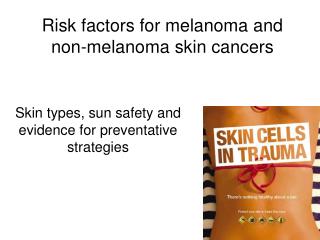
Fair skin + Family history. Prior sun exposure. Blistering sunburns ... Any tan is a sign of damage to the skin. Trying to get a

E N D
Slide 1:Melanoma: What you need to know
Carrine A. Burns, MD Dermatology Associates October 27, 2008
Slide 2:Melanoma
Becoming more common Incidence more than tripled from 1980-2003 Incidence increasing fastest in men age>55 Incidence also rising in children Lifetime risk = 1 in 60 62,480 cases in 2008 Mortality rates stable or decreasing since 1992 8450 fatalities in 2008
Slide 3:Melanoma: What is it?
Cancer of melanocytes �pigment cells� Found normally in skin Increased numbers in moles Mutation (damage to DNA) in the melanocyte leads to uncontrolled growth
Slide 4:Melanoma
Risk factors Fair skin + Family history Prior sun exposure Blistering sunburns Atypical moles History of prior melanoma Tanning booths
Slide 5:Tanning and Melanoma
Strong association between tanning bed use and melanoma Across all ages, 15% higher risk of melanoma 1st tanning bed use at under age 35 leads to 75% increased risk of melanoma Tanning also increases risk of other skin cancer
Slide 6:Tanning = Damage
There is no such thing as a safe tan Any tan is a sign of damage to the skin Trying to get a �base tan� prior to a trip is unwise Self tanners or spray on tans are safe but do not provide any sun protection
Slide 7:Melanoma
Multiple subtypes Not all melanomas are dark Childhood melanoma may more often be red or pink (amelanotic melanoma) Symptoms - often late Itching, Bleeding, Ulceration
Slide 8:Melanoma
Needs to be caught early Horizontal phase (�spreading out�) before vertical growth phase (�growing in�) Months to years Can spread to lymph nodes and other organs Prognosis depends on depth of invasion In Situ (only in top layer of skin) - 100% cure rate >4mm invasion into skin-<50% 5 yr survival
Slide 9:How to detect early?
Patient awareness Perform self skin examinations Physician awareness Full body screening of all patients at risk Prevention is key!!
What to look for: The ABCDE criteriaSlide 18:The Ugly Duckling sign
Slide 19:Melanoma mimics
Lots of things can look like a melanoma but aren�t Still need to be seen and diagnosed by a physician
Slide 20:Normal moles
Nevus=mole Develops during childhood or early adulthood Groups of melanocytes Flat at first Tan or brown Smooth borders Most common on trunk
Slide 21:Normal moles
With time moles change Lose pigment Tan, brown, pink, skin colored Become raised Often end up dome shaped Can have increased hairs
Slide 22:Congenital nevus
�Birthmarks� May be small or large Become raised and bumpy Often have increased hairs Need to be watched for changes Need to be removed if changing significantly
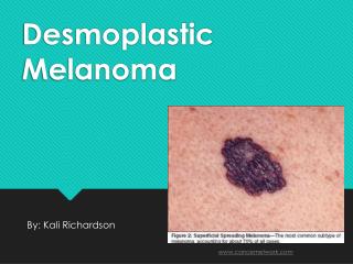
Slide 25:Dysplastic Nevus
Abnormal mole Atypical nevus Potential melanoma precursor 30% of melanomas develop from atypical moles Marker of increased risk Flat, some have raised center Often > 5mm Irregular border, shape, or color pattern Biopsy of suspicious lesions and frequent skin exams
Slide 29:Spitz Nevus
Aka �Juvenile Melanoma� Is NOT malignant Uncommon Can be pink, red or pigmented Complete excision recommended Can be difficult to differentiate from melanoma clinically and histologically
Slide 30:Non-Melanoma Skin Cancer
Much more common than melanoma Basal cell carcinoma Most common form of skin cancer Squamous cell carcinoma Due to sun exposure Rarely life threatening SCC can occasionally spread to lymph nodes Usually just needs local treatment Mohs surgery, excision, scraping and burning, freezing
Slide 31:Basal Cell carcinoma
Slide 32:Squamous Cell Carcinoma
Slide 33:Melanoma: Treatment
Excision In Situ � 0.5cm margin Depth < 1mm � 1cm margin Depth > 1mm � 2cm margin Lymph node evaluation Not needed if depth < 1mm and no other high risk factors Sentinel Lymph Node biopsy Uses dye and radioactivity to find lymph node that melanoma would most likely spread to > 1mm Breslow Gives prognostic information Helps decide who needs more treatment No proven survival benefit Trials ongoing Therapeutic lymph node dissection Removes all the lymph nodes
Slide 34:Melanoma: Further treatments
For patients with spread to other organs or those at high risk No proven effective chemotherapy Interferon alfa-2b FDA approved Evidence mixed Significant side effects Numerous ongoing trials Chemotherapy Vaccine therapy
Slide 35:So what can you do???
Slide 36:PREVENTION AND EARLY DETECTION!!
Slide 38:Melanoma Prevention
There is no such thing as a �healthy tan� Tanning booths cause skin cancer In Maine age<18 must have written permission from parent/guardian to use tanning booth Educate parents Law is often ignored Education around time of proms and events
Slide 39:Melanoma Prevention
Sun Smart Avoid sun when you are able Especially 10am-4pm Wear protective clothing Hats, sunglasses, shirts Sunscreen DAILY!!! for face and hands (SPF15 or >) SPF 30 or 45 for outdoor activities Reapply every 2 � 3 hours or after swimming or sweating Sprays are good for active people
Slide 40:Melanoma: Early Detection
Self Skin examination At least once a month Use mirrors Have someone else check your back Get used to what is there, stable, and normal See a physician for anything new or changing Skin screening by physician Dermatologist if high risk