Download
1 / 26
320 likes | 990 Views
Contact details. (412)-648-6478patersond@dom.pitt.edu. Outline. Classification of pneumoniaEpidemiology

E N D
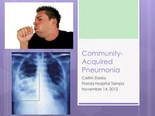
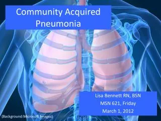
1. Community and Hospital-acquired Pneumonia David L. Paterson MD
Division of Infectious Diseases
2. Contact details (412)-648-6478
patersond@dom.pitt.edu
3. Outline Classification of pneumonia
Epidemiology � who gets pneumonia
Pathogenesis
Causative organisms
Symptoms, signs and investigations
Antibiotic treatment
4. Definition Pneumonia is infection of the gas exchanging (alveolar) compartment of the lung (that is, it is a lower respiratory tract infection)
(Bronchitis is infection of the bronchial tree)
(Tracheitis or pharyngitis are infections of the trachea or pharynx respectively)
5. Importance �Pneumonia is captain of the men of death�
�Pneumonia is the old man�s friend�
Industrialized countries � 3rd leading cause of death
Estimated costs of $7 billion annually
6. Developing countries Pneumonia is a major cause of death in developing countries
Neonatal pneumonia
Pneumonia complicating childhood measles
Pneumococcal pneumonia
Pneumonia complicating HIV infection
7. Classification of pneumonia Why classify pneumonia?
Different categories of pneumonia have different etiologies
Pneumonia is usually treated empirically (that is, before the etiology is known)
Different potential etiologies require different empiric treatments
8. Classification Pneumonia in immunocompetent patients
Community-acquired pneumonia
Hospital-acquired pneumonia (also called nosocomial pneumonia)
Pneumonia in immunocompromised patients
9. Community-acquired pneumonia Frequently referred to as CAP
CAP is pneumonia acquired outside of hospital in the immuno-competent host
Does not include patients with aspiration pneumonia, bronchial obstruction or TB
Nursing home patients
Only 20% of patients with CAP require hospitalization
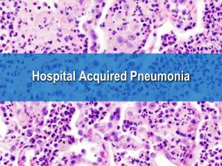
10. Hospital-acquired pneumonia Frequently referred to as HAP
May also be referred to as nosocomial pneumonia
HAP is pneumonia that occurs at least 48 hours after admission to hospital (earlier cases are initially misdiagnosed CAP)
Is the second most common hospital-acquired infection
11. HAP in the ICU Pneumonia acquired in the intensive care unit (ICU) requires special consideration
Many patients in ICU are mechnically ventilated
These patients may develop ventilator-associated pneumonia (VAP)
12. Pneumonia in the immuno-compromised host Differentiated from CAP and HAP because the pathogens are different (however, immuno-compromised patients may also get the pathogens that occur in immuno-competent patients)
Examples of immuno-compromise include HIV, iatrogenic immunosuppression (eg, in transplant recipients) and inherited disorders of the immune system
14. Pathogenesis of pneumonia Defense mechanisms for the lung
How are these defense mechanisms affected?
15. Where is sterile, where is not Bacteria are found in the mouth, nose and pharynx (that is, above the vocal cords)
Examples of the �normal respiratory flora� are viridans streptococci, �non-pathogenic� Neisseria and certain anaerobes
The host defenses prevent these bacteria and others from invading the lower respiratory tree
16. Upper airway defenses Filtering via the nasopharynx
Sneeze reflex
Nasal secretions contain defensins, lysozyme and immunoglobulin A
Glottic closure
Cough reflex
17. Airway protection Mucus layer in trachea and bronchi
Mucociliary escalator
18. Alveolar protection Surfactant proteins
Alveolar macrophages
Neutrophils recruited from vasculature
19. Defects in host defense Impaired cough reflex
Neurologic impairment
Impaired mucociliary function
Smoking
Impaired activity of alveolar macrophages
Corticosteroid use
20. Hematogenous seeding Most infections in the lung are acquired via inhalation or aspiration
Infection of the right sided heart valves (eg, tricuspid valve) can lead to septic pulmonary emboli
22. CAP - Etiology Streptococcus pneumoniae (�the pneumococcus�) is the most common organism causing CAP
Gram positive coccus, often appearing in pairs (�diplococci�)
23. The father of the Gram stain? Christian Gram
24. CAP � Etiology (II) Other common organisms
Haemophilus influenzae
Moraxella catarrhalis
25. CAP � Etiology (III) �Atypical� organisms
Will not grow on conventional culture media
Mycoplasma pneumoniae
Chlamydia pneumoniae
Legionella pneumophila
26. CAP � Etiology (IV) Respiratory Viruses
Metapneumovirus
Influenza
Adenovirus
Parainfluenza viruses
Respiratory syncytial viruses (RSV)
27. CAP- Etiology (V) Less common bacterial causes of CAP
Staphylococcus aureus
Particularly following influenza
Enteric Gram negative bacilli
Klebsiella pneumoniae was a classical cause of CAP but is now rarely seen in the U.S.
Pseudomonas aeruginosa
Usually in severe pneumonia in patients with underlying structural lung disease
28. Important notes In many patients, no etiology is discovered
Prior antibiotic therapy
Autolysis of the pneumococcus
Insufficient testing � atypical agents, viruses etc
29. Clinical pointers to the etiology Young people with no comorbidities and mild disease
Mycoplasma pneumoniae
Young people with no comorbidities and severe disease
Streptococcus pneumoniae
Smokers
Haemophilus influenzae
30. In every case of CAP Antibiotics need to cover:
Streptococcus pneumoniae
Atypical organisms
31. Symptoms of CAP Classical symptoms
Cough (usually productive of purulent sputum)
Fever
Shortness of breath
Pleuritic chest pain
Pneumococcal pneumonia classically starts with shaking chills and patients end up with rusty colored sputum
32. Signs of CAP Patients typically appear short of breath at rest
Vital signs � fever, increased heart rate and increased respiratory rate
Chest exam � crackles over the affected area
Signs of consolidation: ? vocal and tactile fremitus, dullness to percussion, bronchial breath sounds, whispering egophony
33. Investigations � to establish that a patient has CAP CXR
Characteristically abnormal in CAP (some early disease may be an exception)
White blood cell count
- ? WBC with left shift (if bacterial)
Sputum
Gram stain
34. Severe lobar pneumonia
35. CAP - Sputum Gram Stain PRO
Non-invasive, inexpensive
Can establish the etiologic diagnosis
CON
At least one third of patients do not produce sputum
May not change empiric therapy
36. CAP � Etiologic diagnosis For patients requiring hospitalization
Sputum culture
Blood cultures
Legionella urinary antigen
(Pneumococcal urinary antigen)
(Serology for Legionella, Mycoplasma, Chlamydia etc)
37. CAP � Decision Pathway Once the diagnosis of CAP has been made, important decisions need to be made
Does the patient require hospitalization?
If yes, does the patient require ICU admission?
What antibiotics should I prescribe?
38. CAP � the Hospitalization Decision Assessment of pre-existing conditions that compromise the safety of home care
Calculation of the pneumonia severity index (PSI)
Patients are stratified into 5 severity classes
Factors such as age, comorbid conditions, vital signs, mental status
Clinical judgment
39. CAP � antibiotic therapy (I) Need to cover both:
Streptococcus pneumoniae
Atypical organisms (Legionella, Mycoplasma, Chlamydia pneumoniae)
40. CAP � antibiotic therapy (II) Strep. Atypicals
pneumoniae
Penicillin Yes No
Cephalosporins Yes No
Macrolides Yes Yes
Ketolides Yes Yes
Quinolones Yes Yes
Tetracyclines Yes Yes
41. CAP � Antibiotic Therapy (III) Why not monotherapy for everybody?
Antibiotic resistance in Streptococcus pneumoniae
�Collateral damage� inflicted by quinolones
42. Resistance in Streptococcus pneumoniae � Pittsburgh 2005 %R
Macrolides 25%
Doxycyline 20%
Penicillin 10%
Ceftriaxone <1%
Levofloxacin <1%
43. CAP � how to choose a regimen? Consult most recent guidelines
Consider whether therapy will be as an outpatient, inpatient or in ICU
Consider prior antibiotic therapy (predicts resistance to itself)
44. CAP � example regimens Previously healthy patient, no comorbidities, no prior antibiotic use, Rx at home
- Doxycycline PO alone
Patient with comorbidities, prior azithromycin use, Rx at home
- Moxifloxacin PO alone
Patient admitted to medical ward, no prior antibiotic use
- Azithromycin IV plus ceftriaxone IV
45. Special considerations Patients with severe CAP admitted to the ICU
This is the only situation in CAP in which Pseudomonas is required to be covered
Risks for Pseuodmonas in severe CAP include structural lung disease (eg, bronchiectasis, with prior antibiotic use)
47. Hospital-acquired pneumonia Etiologic organisms
Diagnostic tests
Antibiotic regimens
48. HAP � etiologic organisms Early-onset VAP
S. pneumoniae, H. influenzae, S. aureus
Late-onset VAP
Pseudomonas aeruginosa, S. aureus, enteric Gram negative bacilli (Klebsiella, E. coli etc)
HAP, not associated with ventilators
Enteric Gram negative bacilli, H. influenzae, S. aureus
49. HAP � symptoms and signs Most patients with HAP are ventilated or neurologically impaired, so can not describe symptoms
Nursing observations
Fever
Increased respiratory rate
Increased oxygen requirement
Suctioning of purulent respiratory secretions
50. HAP � laboratory findings WBC
? WBC with left shift
CXR
Alveolar infiltrate
BUT
- Could also be due to pulmonary edema, blood, ARDS etc
51. VAP � making the diagnosis Two approaches
Standardized clinical approach
Clinical pulmonary infection score (CPIS)
Microbiologic approach
Quantitative cultures of LOWER respiratory secretions obtained by bronchoalveolar lavage (BAL) or protected specimen brush (PSB)
52. Colonization versus pneumonia Ventilated patients routinely have colonization of the airways with potentially pathogenic bacteria
There is no advantage in treating colonizing bacteria in the absence of pneumonia
Excessive antibiotic use in the ICU leads to increased antibiotic resistance
53. Empiric antibiotic therapy � HAP (I) Early onset VAP
Ampicillin/sulbactam
Rationale: covers S. pneumoniae, H. influenzae, S. aureus
54. Empiric antibiotics � HAP (II) Late-onset VAP
Combination of vancomycin, cefepime and gentamicin
Rationale: Vancomycin in case there is MRSA, cefepime and gentamicin in case there is multiply resistant Pseudomonas
55. Late-onset VAP EMPIRIC therapy should usually consist of combination therapy because antibiotic resistance is common in bacteria in the ICU
Therapy should then be narrowed when antibiotic susceptibilties are known
56. Empiric therapy � HAP (III) HAP in the non-ventilated patient
Empiric therapy: Piperacillin/tazobactam
Rationale � covers common Gram negative bacilli, anaerobes
57. SUMMARY Pneumonia can be subdivided into CAP, HAP and pneumonia in the immunocompromised
The subdivisions are important because different organisms are responsible
Pneumonia treatment is almost always empiric, therefore knowledge of the likely pathogens is essential
58. Practice question 1 Which of the following are not true with respect to CAP:
Less than 50% of patients with CAP require admission to hospital
Treatment of CAP with ceftriaxone and azithromycin is logical because it provides coverage of pneumococci and atypicals
Knowledge of the prior antibiotic treatment of a patient with CAP is important because it will modify the empiric regimen chosen
Coverage of MRSA and Pseudomonas are important in all cases of CAP
59. Practice question 2. With respect to VAP, which of the following is not true:
Since patients with VAP are in the ICU, multidrug resistance is common
Antibiotic treatment for VAP can not wait until the susceptibilties of the infecting organisms are known
Pulmonary edema or ARDS may be hard to distinguish radiologically from VAP
Pneumocystis carinii should always be covered in an antibiotic regimen for VAP