Unit-1 Respiration
What is Respiration? Respiration is a metabolic process that occurs in all organisms. It is a biochemical process that occurs within the cells of organisms. In this process, the energy (ATP-Adenosine triphosphate) is produced by the breakdown of glucose which is further used by cells to perform various functions.


Unit-1 Respiration
E N D
Presentation Transcript
Shri Shivaji Education Society, Amravati'sSHRI PUNDLIK MAHARAJ MAHAVIDYALAYA, NANDURA RLY. DIST. BULDANA Topic: Unit-I: Respiration Class: B.Sc-III, Sem-V Mr. Shantaram Bhoye Assistant Professor & Head M.Sc., NET-JRF, SET, M.A.(Eng.)
Respiration: • Respiration is a physiological process occurring in all living organisms. • Every cell requires energy for carrying out its metabolic activities. • The cell consumes oxygen during oxidation of substances resulting in the release of heat and energy and production of carbon dioxide. • The carbon dioxide acts as a poison for protoplasm unless removed from the body. • The system designed for exchange of gases between the organism and its environment is termed the respiratory system.
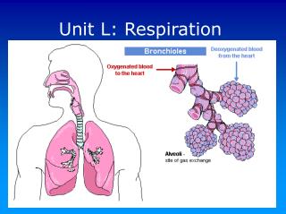
Structure of respiratory organs: Gills and Lungs • Exchange of oxygen and carbon dioxide in an organism takes place at two locations. • During internal respiration gaseous exchange occurs between blood and cells of the. • During external respiration, the gaseous exchange takes place between blood and the external environment. • Blood serves as transport medium for carrying oxygen to the body cells and carbon dioxide away from the body cells. • The body structures which are needed for gaseous exchange between the blood and the surrounding medium are known as respiratory organs. • Depending on the type of medium, vertebrates have two principal types of respiratory organs- gills (for aquatic respiration) and lungs (for terrestrial respiration).
A. Gills: Gills or branchia are the aquatic respiratory organs of molluscs, many crustaceans, fishes and amphibians. In addition to gas exchange, gills may serve for loss or gain of water and elimination of salts in marine teleost's. On the basis of their location, gills are of two types-internal gills and external gills. In some animals both internal and external gills are present. I. Internal gills or true gills: The gills are characteristics of fishes. In chordate embryo the pharyngeal cavity is connected to the outside by a series of lateral openings called, gill slits or pharyngeal clefts. The number of gill slits varies in different chordates- 140 pairs in Amphioxus, 6-14 pairs in cyclostomes, 5 pairs in most elasmobranchs and bony fishes, and 4 pairs in teleosts.
Structure of a true gill: • They are supported by branchial cartilage. • A single row of lamellae on one side of branchial septum forms half gill called a demibranch or hemibranch. • A branchial septum with two attached demibranchs forms a complete gill or holobranch. • The gill filaments are richly supplied with blood capillaries. • On the surface of gill filaments exchange of gases through water takes place. • In bony fishes a bony flap called operculum covers the gills.
II. External or larval gills: • They are ectodermal in origin and are usually temporary organs found only in larval stages, therefore they are also called larval gills. • They occur in the larvae of Lampreys, a few bony fishes, lung fishes and all amphibians. • In amphibians, larval gills are absorbed at the time of metamorphosis. • In some urodels both external gills and gill slits persist during adult life. • In amphibia gills are absorbed but gill slits persist. • Gills assume different shapes: pectinate, bipinnate, dendritic, leaf like, etc. • Each gill consists of a narrow main central axis bearing a double row of filaments. • They are vascularized by aortic arches.
B. Lungs: • Lungs are the respiratory organs of land vertebrates and lung fishes. • They are very elastic and distensible. • In vertebrate embryos, lungs developed as a single midventral diverticulum (lung primordium) from the floor of the pharynx. • It soon bifurcates into right and left lung buds. • The undivided common portion develops into windpipe called trachea and larynx which opens into pharynx through glottis. • Each lung has an inner endodermal lining derived from embryonic gut, an outer visceral peritoneum and in between the two, mesodermal mesenchyme containing lymph and blood vessels, nerves, smooth muscle fibers and connective tissues.
A) The lungs of modern amphibia: • They are simple hollow sacs a wide central cavity. • They are elongated in urodels but bulbous in anurans. • Left lung is usually longer in urodels but rudimentary in caecilians. • In frogs and toads lung wall may be divided peripherally by network of folds or trabeculae into air sacs or alveoli. • They are richly vascular.
B) Lungs of Reptiles: • Lungs are reptiles are more complicated. • In Sphenodon and snakes, lungs are simple thin walled sacs. • In legless lizards and snakes left lung may be rudimentary or absent. • In lizards and turtles, wall of lung is greatly thick and spongy. • In Chamaeleon several air sacs arise from the distal portion of lungs.
C) Lungs of Aves (Birds): • Avian lungs are unique in architecture and greatly modified due to their arial mode of life. • Lungs in birds are small, compact, spongy and have very little capacity of contraction and expansion. • They have several thin walled, membranous air sacs. • The bronchus divides repeatedly forming a network of air capillaries which do not terminate blindly. • Due to this the avian lungs become highly efficient organs.
D) Lungs of Mammals: • They are highly developed, spongy and very elastics. • They lie in pleural cavities. • In mammals, externally the lungs are subdivided into lobes. • Usually there are only two left lobes. • Right lung has 3 lobes in Man and 4 lobes in Rabbit. • In some mammals, such as whales, elephant, etc. lungs are simple and without lobes. • In monotremes and rats only the right lung is lobulated. • The bronchus divides repeatedly inside the lung and ultimately terminate in alveoli. • They are terminal and blind they always retain certain amount of residual air after every expiration. • In mammals, intercoastal muscles, ribs, diaphragm, sternum and abdominal muscles, all help in breathing.
REGULATION OF VENTILATION IN LUNGS: • During normal quite breathing, inspiration is the active process involving contraction of diaphragm and external intercoastal muscles, whereas, expiration is the passive process involving recoiling of lungs and thoracic cage. • The process of breathing or ventilation of lungs takes place in two distinct steps: • Inspiration or breathing air into the lungs. • Expiration or expelling out air from the lungs. • The movement of air in and out of the lungs occurs because of alternated changes in intra-pulmonary pressure which is brought about both during inspiration and expiration by the contraction and relaxation of diaphragm and the intercoastal muscles. Mr. Shantaram Bhoye, Assistant Professor, Shri Pundlik Maharaj Mahavidyalaya, Nandura Rly., Dist. Buldana
A) Mechanism of Inspiration: • During inspiration the size of thoracic cavity increases in all directions. • The enlargement is brought about by the flattening of diaphragm and lifting up of the thoracic basket. • The diaphragm is formed of a thin sheet of radial muscles present on the floor of thoracic cavity. • In resting position it is dome-shaped. • During inspiration, when the radial muscles contract, diaphragm flattens and descends down into the abdominal cavity. • The ribs are provided with two sets of muscles: external intercostal muscles and internal intercostal muscles. • When thoracic cavity increases in size, the pleural cavities expand and negative pressure is increased in it. • Now lungs enjoy greater space and expand. Mr. Shantaram Bhoye, Assistant Professor, Shri Pundlik Maharaj Mahavidyalaya, Nandura Rly., Dist. Buldana
Mechanism of expiration: • During expiration, changes occur in opposite direction. • Due to relaxation diaphragm and ribs are brought to their original position and form. • The diaphragm assumes dome-shaped appearance. • Therefore, volume of thoracic cavity and also the negative pressure of pleural cavities is reduced. • This exerts pressure on lungs and the air filled in the lungs is compressed and is squeezed out. • Thus, the expiratory phase is a passive event in breathing. Mr. Shantaram Bhoye, Assistant Professor, Shri Pundlik Maharaj Mahavidyalaya, Nandura Rly., Dist. Buldana
Thoracic cage and diaphragm play in respiration varies from animal to animal and from year to year in the same animal. • The lungs are not totally emptied by any movement of breathing and some air is left in air passages. • Therefore, air is never changed completely during inspiration but is merely freshened up. Mr. Shantaram Bhoye, Assistant Professor, Shri Pundlik Maharaj Mahavidyalaya, Nandura Rly., Dist. Buldana
Neurophysiologic control of respiration: • Respiration is an involuntary process and is carried out automatically at a constant rate under normal conditions. • The contraction of various respiratory muscles at proper time and with proper strength to secure adequate gaseous exchange. Nervous Control: • Medullary respiratory center (The vital knot of Fluorenes): • It is situated in the floor of 4th ventricle present in the medulla. • The center is bilateral and two halves are connected together by commissural neurons. • The neuron cells of the center are connected to the breathing apparatus with motor as well as sensory (afferent and efferent) nerves and thus maintain a reflex arch. • These cells are sensitive to changes in chemical composition of blood and concentration of Carbon dioxide in plasma. Mr. Shantaram Bhoye, Assistant Professor, Shri Pundlik Maharaj Mahavidyalaya, Nandura Rly., Dist. Buldana
Each half of the respiratory center is composed of two parts: Inspiratory and Expiratory center. • The expiratory center lies above the inspiratory center. • Out of the two, only one works at a time. • According to Pitts, Magoun and Ranson, the inspiratory center works in normal breathing and expiratory center during forced expiratory conditions like sneezing, laughing and coughing. • In the wall of alveoli of lungs are present stretch receptors or baroreceptors. • These are stimulated by the expansion and relaxation of lungs. • These receptors propagate inhibitory impulses to the inspiratory and expiratory parts of the respiratory center respectively. • Pneumotaxic center lies in the upper part of pons. It maintains rhythmicity of respiration. • It is connected with both inspiratory and expiratory centers. Mr. Shantaram Bhoye, Assistant Professor, Shri Pundlik Maharaj Mahavidyalaya, Nandura Rly., Dist. Buldana
Hering-Breuer Reflexes: • The respiratory center is connected with baroreceptors or stretch receptors present in the lungs and with the chemoreceptors located in the carotid sinuses and aortic arch through afferent nerve fibers. • The inspiratory center propagates motor impulse spontaneously to the external intercostal muscles and the muscles of diaphragm and also the pneumotaxic center. • As a result these inspiratory muscles contract and the thorax and lungs expand, filling the lungs with fresh air. • This stimulates baroreceptors in the lungs. • These send inhibitory impulses to inspiratory center, resulting in the relaxation of inspiratory muscles and expiration follows by the initiation of Hering-Breuer expiratory reflex. • When expiration is completed, the lungs are deflated and lung baroreceptors are inhibited to start Hering-Breuer inspiratory reflex. Mr. Shantaram Bhoye, Assistant Professor, Shri Pundlik Maharaj Mahavidyalaya, Nandura Rly., Dist. Buldana
Chemical Control: • Rate of breathing is controlled by Carbon dioxide level of the arterial blood and cerebrospinal fluid. • Chemoreceptors in the brain, aortic arch and carotid sinus detect carbon dioxide, pH and Oxygen levels in blood and conduct the information to the brains rhythmic center. • Carbon dioxide has more effect on breathing than the level of oxygen. • When Carbon dioxide content of blood drops below a certain critical level, breathing stops and with the increase in Carbon dioxide level in blood, breathing is accelerated. • By repeated deep inhalations and exhalations in rapid succession, Carbon dioxide level in blood falls and breathing becomes normal. Mr. Shantaram Bhoye, Assistant Professor, Shri Pundlik Maharaj Mahavidyalaya, Nandura Rly., Dist. Buldana
Respiratory pigments: • The uptake, transport and delivery of Oxygen and Carbon dioxide is done by a group of colored proteins (chromoproteins) called respiratory pigments. • These pigments are capable of forming a loose, reversible combination (Oxyhaemoglobin) with O2 (sometimes with CO2) when exposed to O2 high tension and releasing O2 readily at lower tensions at level of tissues. • There are different pigments in different phyla and even several different pigments in the same phylum or more than one pigment exists in the same animal. • Respiratory pigments are present in RBCs or in different components of blood. • Those pigments present outside the blood cells (extracellular fluid) do not function as carriers of O2 and CO2. Mr. Shantaram Bhoye, Assistant Professor, Shri Pundlik Maharaj Mahavidyalaya, Nandura Rly., Dist. Buldana
1. Haemoglobin (Hb): • It is the most familiar, most efficient respiratory pigment. • It occurs in majority of vertebrates and invertebrates. • In invertebrates it is found dissolved in plasma, whereas in vertebrates it its contained in the special cells, called red blood corpuscles (RBC). • Its Oxygen carrying capacity is more than other respiratory pigments. • It is an iron containing compound and consists of an iron-porphyrin compound (heme) associated with a protein globin. • Heme is a metalloporphyrin. • Molecular weight of Hb in RBC 68,000 Dalton. • In human beings each gram of Hb can combine with 1.3 ml of O2 in normal man. • 100 ml of blood of normal man contains about 16 gms of Hb and in normal woman it is about 14 gms. • At the level of respiratory surface Hb combines with O2 to form oxyhemoglobin while at tissue level O2 is released. • Hb also carries some CO2 in the form of carbhaemoglobin from tissues to the lung alveoli for its removal to exterior. Mr. Shantaram Bhoye, Assistant Professor, Shri Pundlik Maharaj Mahavidyalaya, Nandura Rly., Dist. Buldana
2. Chlorocruorin: • It is the second major respiratory pigment found in blood of invertebrates. • It is distributed in only four annelid families of the class polychaeta. • It occurs only in plasma. • The affinity of chlorocruorin for O2 is equivalent to Hb. • It is within the same family of worms like sabellidae, serpulidae and amphotritidae some species have chlorocruorin while other have Hb. • In serpula both these pigments are present. • The oxygen carrying capacity of chlorocruorin is 9.1 ml pf O2/100 ml of blood. • As compared to Hb it has higher affinity for CO2. • In dilute state it is green in colour while in concentrated form it is red in color. Mr. Shantaram Bhoye, Assistant Professor, Shri Pundlik Maharaj Mahavidyalaya, Nandura Rly., Dist. Buldana
3. Hemocyanin: • Hemocyanin is a copper-containing pigment without any porphyrin group. • It always occurs dissolved in plasma. • It is the main respiratory pigment occurring in two important phyla i.e. Mollusca and Arthropoda. • It is colourless when deoxygenated and blue when oxygenated. • It is transport O2. Mr. Shantaram Bhoye, Assistant Professor, Shri Pundlik Maharaj Mahavidyalaya, Nandura Rly., Dist. Buldana
4. Haemerythrin: • Haemerythrin is a iron containing blood pigment in which iron is directly attached to protein, as porphyrin is absent. • It is found in marine invertebrates from sipunculeds, Priapulids, Brachiopods and in Annelid worm Magalona. • Pigment appears purple when oxygenated and becomes colourless when reduced. • It is a protein with M.W. 1,08,000. • Vascular haemerythrin transports O2 from high O2 pressure of sea water to the coelom and coelomic haemerythrin and thereby constitute a transfer system between vascular pigments and tissue. Mr. Shantaram Bhoye, Assistant Professor, Shri Pundlik Maharaj Mahavidyalaya, Nandura Rly., Dist. Buldana
Transport of Respiratory gases: Transport of Oxygen: 1. In simple solution: • Oxygen dissolves in water of plasma and is transported in physical form. • The amount of oxygen transported in this way is very negligible only 0.3 ml/100 ml of plasma is transported in this way. • This is because of poor solubility of oxygen in water content of plasma. • Still the transport of oxygen in this form becomes important during excess demand of oxygen by the tissues (muscular exercise). Mr. Shantaram Bhoye, Assistant Professor, Shri Pundlik Maharaj Mahavidyalaya, Nandura Rly., Dist. Buldana
2. In combination with Hb: • Oxygen combines with Hb to form oxyhemoglobin. • 97% of oxygen is transport by this method. • When the partial pressure of oxygen in the blood is more, Hb combines readily with oxygen and wherever the partial pressure of oxygen in blood is less, Hb releases oxygen. • Each molecule of Hb contains 4 atoms of iron. • Each iron atom combines with one molecule of oxygen. • The combination of oxygen with Hb is called oxygenation because iron remains in ferrous form only. • Hb4 + 4O2↔Hb4 O8 • The amount of O2 transported by blood is called oxygen carrying capacity of blood. • One gram of Hb carries 1.34 ml of oxygen. Mr. Shantaram Bhoye, Assistant Professor, Shri Pundlik Maharaj Mahavidyalaya, Nandura Rly., Dist. Buldana
Oxygen Hb dissociation curve: • This curve demonstrates the relationship between partial pressure of Oxygen and the percentage saturation of Hb with oxygen. • It explains affinity of Hb for oxygen. • Normally, Hb is saturated with oxygen only up to 95%. • This saturation of Hb with oxygen depends upon the partial pressure of oxygen. • When the partial pressure of oxygen is more, Hb accepts oxygen and when partial pressure of oxygen is less, Hb releases oxygen. Mr. Shantaram Bhoye, Assistant Professor, Shri Pundlik Maharaj Mahavidyalaya, Nandura Rly., Dist. Buldana
The partial pressure of oxygen and saturation of Hb are plotted to obtain the oxygen Hb dissociation curve. • Under normal conditions, oxygen Hb dissociation curve is ‘S’ shaped or sigmoid shaped. • The lower part of the curve indicates dissociation of oxygen from Hb. • The upper part indicates the acceptance of oxygen by Hb, depending upon the partial pressure of oxygen. • PO50 is the partial pressure of oxygen at which Hb saturation with oxygen is 50%. Mr. Shantaram Bhoye, Assistant Professor, Shri Pundlik Maharaj Mahavidyalaya, Nandura Rly., Dist. Buldana
The oxygen Hb dissociation curve is shifted to left or right by various factors. 1. Shift to left indicates acceptance of Oxygen by Hb. 2. Shift to right indicates dissociation of Oxygen by Hb. Mr. Shantaram Bhoye, Assistant Professor, Shri Pundlik Maharaj Mahavidyalaya, Nandura Rly., Dist. Buldana
Bohr’s Effect: • It is the effect by which the presence of carbon dioxide, decreases the affinity of Hb for oxygen. • It was postulated by Christian Bohr in 1904. • Due to continuous metabolic activities the partial pressure of Carbon dioxide increases in the tissue and partial pressure of oxygen decreases in the tissue. • Due to this Carbon dioxide enters the blood and oxygen is released from the blood to the tissue. • If enhances further release of oxygen to the tissue and the oxygen dissociation curve is shifted to right. This is due to Bohr’s effect. Mr. Shantaram Bhoye, Assistant Professor, Shri Pundlik Maharaj Mahavidyalaya, Nandura Rly., Dist. Buldana
Transport of Carbon dioxide: • Carbon dioxide transported by the blood from tissues to the alveoli. • Carbon dioxide is transported in the blood in four ways- • As dissolved form- 7% • As carbonic acid- negligible • As bicarbonates- 63% • As carbamino compounds- 30% 1. Transport of Carbon dioxide as dissolved form: • Carbon diffuses into blood and dissolves in plasma forming a simple solution. • Only about 3 ml/100 ml of plasma of carbon dioxide is transported in dissolved form. 2. Transport of Carbon dioxide as carbonic acid: • Part of dissolved carbon dioxide in plasma combines with the water to form carbonic acid. • This reaction is very slow and a negligible amount of carbon dioxide is transported in this form. Mr. Shantaram Bhoye, Assistant Professor, Shri Pundlik Maharaj Mahavidyalaya, Nandura Rly., Dist. Buldana
3. Transport of Carbon dioxide as Bicarbonate: • About 63% of carbon dioxide is transported as bicarbonate. • From plasma, carbon dioxide enters the RBCs. • In the RBCs, CO2 combines with water to form carbonic acid under the action of an enzyme called carbonic unhydrase. • It dissociates into bicarbonate and hydrogen ions. • The concentration of bicarbonate increases more and more in the cell which causes its diffusion through the cell membrane of RBCs into the plasma. Mr. Shantaram Bhoye, Assistant Professor, Shri Pundlik Maharaj Mahavidyalaya, Nandura Rly., Dist. Buldana
Chloride shift or Hamburger phenomenon: • The exchange of chloride ion for a bicarbonate ion across the cell membrane of RBC is called chloride shift. • It was discovered by Hartog Jakob Hamburger in 1892. • Chloride shift occurs when CO2 enters the blood from the tissues. • Plenty of sodium chloride is present in plasma. • It dissociates into sodium and chloride ions when the negatively charged bicarbonate ions move out of RBC into the plasma, the negatively charged chloride ions move into the RBC in order to maintain the ionic balance. • The bicarbonate ions combine with sodium ions in the plasma and form sodium bicarbonate which is transported by the blood. • The hydrogen ions dissociates from carbonic acid are buffered by Hb inside the RBC. Mr. Shantaram Bhoye, Assistant Professor, Shri Pundlik Maharaj Mahavidyalaya, Nandura Rly., Dist. Buldana
Reverse chloride shift: • The process by which the chloride ions are moved back into plasma from RBCs is called reverse chloride shift. • When the blood reaches the alveoli, NaHCO3 dissociates into NA+ and HCO3 ions. • Bicarbonate ion moves into RBC, which makes Cl- ion to move out of the RBC into the plasma, where it combines with sodium and forms sodium chloride. • At the same time, O2 also enters the RBC. • It displace H+ from Hb and combines with HCO3 to form H2CO3 which dissociates into H2O and CO2. Then the CO2 is expelled out. Mr. Shantaram Bhoye, Assistant Professor, Shri Pundlik Maharaj Mahavidyalaya, Nandura Rly., Dist. Buldana
4. Transport of CO2 as Carbamino compounds: • About 30% of CO2 combines with Hb to from carbamino-haemoglobin or carbhaemoglobin and it combines with plasma proteins to form carbamino-proteins. • carbamino-haemoglobin and carbamino-protein are together called carbamino compounds. • CO2 combines with Hb or proteins with a loose bond so that, CO2 is easily release into alveoli, where partial pressure of CO2 is low. Mr. Shantaram Bhoye, Assistant Professor, Shri Pundlik Maharaj Mahavidyalaya, Nandura Rly., Dist. Buldana