Craniometry and Functional Craniology
Craniometry and Functional Craniology. Part II: Functional Craniology: Kinematics and Dynamics. Michael S. Yuan, DDS, MA, PhD Assistant Professor of Clinical Dentistry. Division of Orthodontics School of Dental and Oral Surgery Columbia University. December 4, 2003. Lecture outline.

Craniometry and Functional Craniology
E N D
Presentation Transcript
Craniometry and Functional Craniology Part II: Functional Craniology: Kinematics and Dynamics Michael S. Yuan, DDS, MA, PhD Assistant Professor of Clinical Dentistry Division of Orthodontics School of Dental and Oral Surgery Columbia University December 4, 2003
Lecture outline 1. Introduction: definition, scope, and objectives 2. Kinematics and dynamics 3. Biomechanics: forces, deformation, stresses, strains 4. Form and Function 5. Bone remodeling and growth directions 6. Moss’ Hypothesis: Functional Matrix Hypothesis 7. Clinical applications
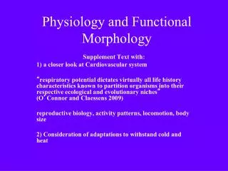
Functional Craniology • Definition: The study of the craniofacial complex in relation to the fields of functional anatomy, comparative anatomy, embryology, and growth and development. • Scope: anatomy, embryology, histology, physiology, growth and development of the head and neck regions; theories of craniofacial growth; craniometry and cephalometry; and others • Objectives: 1) to relate the function to the morphology of the craniofacial complex. 2) to apply the theories of craniofacial growth and biomechanics to better understand the morphology, ontogeny and phylogeny of the craniofacial complex 3) to provide the scientific basis for the clinical applications in the treatment of craniofacial anomalies.
Kinematics The measurement and description of the changes in size, shape, and location of the craniofacial complex. Dynamics The interpretation and description of the biological processes of the changes in size, shape, and location of the craniofacial complex.
Kinematics The description of measurement. The description of the changes in size, shape, and location based on observations and measurements. The why, who, how, which and where, and what in measurement. 1) The history, scope, definition, and objectives of anthropometry 2) Introduction to craniometry and cephalometry 3) Define anatomical landmarks 4) Define anthropometric, craniometric, cephalometric measurements 5) Measuring devices and technical assessments 6) Data analysis, result descriptions a) qualitative vs quantitative b) absolute vs relative c) statisticalanalysis
Dynamics What is the true meaning of a measurement? How to see beyond the numbers? And what are we looking for? What makes the changes in size, shape, and location of an organism or a structure (the transformation)? What are the modern hypotheses, paradigms, and syntheses in understanding these kinematic changes? 1) Introduction to functional craniometry 2) Basic principles in growth and development, especially in osteology and biomechanics. 3) The functional, biological, and mechanical interpretations of the transformation of an organism or a structure. 4) The evolutionary significance: the adaptation and the selection 5) Clinical applications
Terminology used in Biomechanics Force: compression, tension, bending, shear, and torsion Deformation: Change of form due to the loading of forces Stress: the force per unit area Strain: the dimensional change expressed as a fraction (ratio) of the subject’s original size
Compression: compression is the direct expression of the force, which pushes everything towards the center of an object. Tension: the opposite of compression; the force which pulls everything away from the center; where there is a compressive force, there must be a tensile force. Shear: shear is present, when two forces are thrusting in opposite directions but offset and slide past each other. Bending: is found between the pulling of tension and the pushing of compression. Torsion: a result of all the other four forces. Torsion is twist. Torsion is actually a specialized bending, a circular bending. Force Compression, Tension, Shear, Bending, Torsion 1) Two basic forces: Compression & Tension 2) A combination of compression and tension: Shear & Bending 3) A combination of the above four forces: Torsion
Tension Original status Compression
Shear Original status Bending
Original status Torsion
Skeletal Class II, convex profile Skeletal Class III, concave profile Source: Dr. Wisanu Charoenkul Source: Dr. Sonia Abraham Facial Deformation
Cephalic Form, Facial Form, and Arch Form Dolichocephalic (long and narrow head) Leptoprosopic (long and narrow face) Dolichuranic (V shape, narrow maxillary arch) Source: Dr. Christel Hummert FM, female,13y 6m
* Mouth breather; Enlarged pharyngeal tonsil (adenoid) FM Female 13y 6m Source: Dr. Christel Hummert
Form (Structure) and Function Form (structure) follows Function. Function determines form (structure). Function controls form (structure). Function regulates form (structure). Form (structure) is the realization of information and the product of the functional attributes.
Cranial Sutures • 1. Edge-to-edge suture • No force loading • 2. Beveled suture • Shear force [Squamosal suture] • 3. Serrated suture • Intermittent tension force [Sagittal suture] • 4. Beveled and serrated suture • Intermittent tension and shear force • 5. Butt-ended sutures • Intermittent compressive force “Form Follows Function”
Synovial Joints (I) • 1. Plane (gliding) joint • Sliding motion of all directions • 2. Hinge joint • Flexion/extension [ Humeroulnar joint] [ Intermetatarsal joint] “Form Follows Function” Illustrations: http://www.science.ubc.ca/~biomania/tutorial/bonejt/intro.htm
Synovial Joints (II) • 4. Ellipsoidal (condyloid) joint • flexion/extension, adduction/abduction, circumduction, but no rotation • 3. Pivot joint • Rotation [Radioulnar joint] [Temporomandibular joint] “Form Follows Function” Illustrations: http://www.science.ubc.ca/~biomania/tutorial/bonejt/intro.htm
Synovial Joints (III) • 6. Ball and socket joint • flexion/extension, • adduction/abduction, • circumduction, and rotation • 5. Saddle joint • Similar to ellipsoidal joint, • but freer [First carpometacarpaljoint] [First carpometacarpal joint] [Glenohumeral joint] [Glenohumeral joint] “Form Follows Function” Illustrations: http://www.science.ubc.ca/~biomania/tutorial/bonejt/intro.htm
6 2, 3 1 4 5 Functional Structure of Skull (From a mechanical point of view) In the force loading areas, pillar-like struts serve as mechanically efficient reinforcements to resist and dissipate pressure and traction, especially to the masticatory force. 1) Fronto-nasal pillar 2) Zygomatic arch pillar with vertical branch 3) Zygomatic arch pillar with horizontal branch 4) Basal arch in upper jaw 5) Basal arch in lower jaw 6) Occipital pillar 7) Pterygoid-palate pillars
a b c e d Functional Structure of Skull (From a mechanical point of view) • In the non- or less force loading areas, adipose tissue and pneumatic cavities fill those mechanically neutral areas. 1) Paranasal sinuses a) Frontal sinus b) Ethmoid sinus c) Sphenoid sinus d) Maxillary sinus 2) Accessory tympanic spaces e) Mastoid air cells
Hominid: Australopithecine Pongid: male gorilla Temporal muscle fibers oriented towards the posterior teeth; emphasis on the posterior teeth in mastication and dietary adaptation Temporal muscle fibers oriented towards the anterior teeth; emphasis on the anterior teeth in mastication and dietary adaptation Sagittal crests and temporal muscle orientations Hominids compared to pongids
Bone remodeling Deposition: the biological process of laying down the bone Resorption: the biological process of removing the bone Remodeling: A basic part of bone growth involves simultaneous deposition and resorption on all inner and outer surfaces of the entire bone. It provides regional changes in shape, dimensions, and proportions. Drift: Growth movement of an enlarging portion of a bone by the remodeling. The combinations of deposition and resorption result in growth movement toward the depository surface. Displacement: The growth movement of a whole bone as a unit. The bone is carried away from its articulation in relation to other bones. Direction of growth: 1) the direction of drift 2) the direction of displacement 3) the net direction of drift and displacement.
The Growth of the Coronoid Process Deposition (+); Resorption (-); Direction of growth (arrow)
The Growth of Mandible Deposition (blue arrow); resorption (white arrow)
Drift vs Displacement Drift: the growth movement of an enlarging portion of a bone by the remodeling. Displacement: The growth movement of a whole bone as a unit. Direction of growth: the net growth direction of drift plus displacement.
Head (craniofacial complex) is a region, where a series of functions are carried out. These functions include vision, hearing, speech, mastication, swallowing & digestion, respiration, neuralintegration, and others. The successful execution of a function requires biomechanical protection and support. Moss’ craniofacial growth theory: Function of the craniofacial complex region is performed by the Functional Cranial Components (F.C.C).
Functional Matrix Hypothesis (Moss’ Hypothesis) “The functional matrix is primary and the presence, size, shape, spatial position, and growth of any skeletal unit is secondary, compensatory, and mechanically obligated to changes in the size, shape, spatial position of its related functional matrix” (Moss, 1968)
Functional Matrix Hypothesis (Moss’ Hypothesis) “The origin, development and maintenance of all skeletal units are secondary, compensatory and mechanically obligatory responses to temporally and operationally prior demands of related functional matrices.”
THE FUNCTIONAL MATRIX HYPOTHESIS One Function Functional Cranial Component Functional Matrix Skeletal Unit 1. Periosteal Matrix -------------------------------> 1. Microskeletal 2. Capsular Matrix --------------------------------> 2. Macroskeletal a. Masses b. Functioning spaces
Types of Functional Matrix 1. Periosteal matrix (e.g., muscles) Active growth Deposition and resorption Affect size and/or shape 2. Capsular matrix (e.g., brain, oral cavity) Passive growth No deposition No resorption Affect location
Craniofacial Growth Growth Active growth process 1) Sutural growth 2) Bone remodeling 3) Cephalic cartilage growth Active growth (Periosteal) + Passive growth (Capsular) = Total growth Passive growth process 1) The growth of neural, orbital, CSF, and other masses and real substances 2) The expansion of oro-naso-pharygeal and other functioning spaces
1. Orthodontics Periosteal Matrix ------------> Skeletal Unit [Teeth] [Alveolar Bone] 2. Dentofacial Orthopedics and Orthognathic Surgery Capsular Matrix -------------> Multiple Skeletal Units [Functional Appliances] [Jaw Bones] Capsular Matrix -------------> Multiple Skeletal Units [Distraction osteogensis: e.g., hemifacial microsomia] [Jaw Bones] 3. Craniofacial surgery Capsular Matrix -------------> Multiple Skeletal Units [Craniotomy: e.g. Crouzon Syndrome] [cranial bones] [Distraction osteogensis: e.g., Treacher Collin Syndrome] [facial and jaw bones] Use of the “Functional Matrix” in the therapy of orthodontics, dentofacial orthopedics, and orthognathic and craniofacial surgery
Introduction: definition, scope, and objectives Kinematics and dynamics Biomechanics: forces, deformation, stresses, strains Form and Function Bone remodeling and growth directions Moss’ Hypothesis: Functional Matrix Hypothesis Clinical applications
References Enlow, D.H. (1990). Handbook of Facial Growth (3rd edition). Philadelphia, Pennsylvania: W.B. Saunders Company. Moyers, R.E. (1988). Handbook of Orthodontics (4th edition). Chicago, Illinois: Year Book Medical Publishers, Inc. Proffit, W.R. (2000). Contemporary Orthodontics (3rd edition). St. Louis, Missouri: Mosby, Inc. Ranly, DM (1980). A Synopsis of Craniofacial Growth. Norwalk, CT: Appleton-Century-Croft.
Acknowledgments Thanks to Professor Melvin L. Moss Professor Letty Moss-Salentijn Professor Alfonso Solimene And Dr. Christel Hummert