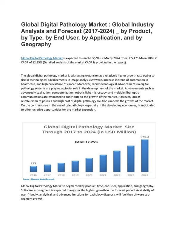
1 / 4
30 likes | 50 Views
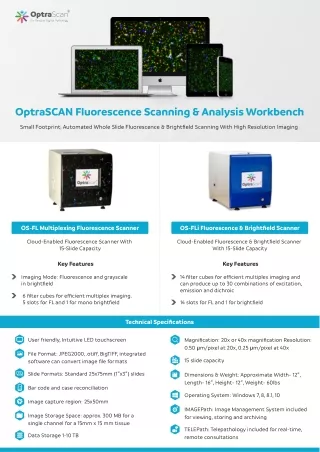
OptraSCAN offers OS-FL fluorescence image slide scanners & OS-FLi fluorescence & brightfield scanners that are cloud-enabled and ideal for fluorescence scanning. It can scan 15 tissue slides at a time. They come with OptraSCANu2019s proprietary software such as IMAGEPathu00ae, the Image Management System apt for viewing, storing, and archiving as well as TELEPathu00ae<br><br>Tel : 1-408-524-5300<br>Contact us at- info@optrascan.com <br>Visit- https://www.optrascan.com/products/os-fl-multiplexing-fluorescence-scanner

E N D
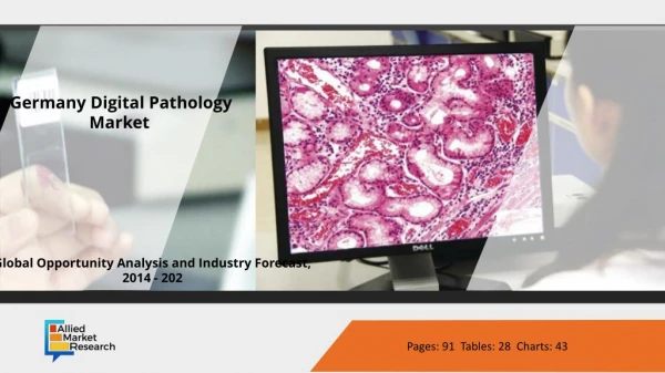
OptraSCANFluorescenceScanning & AnalysisWorkbench SmallFootprint, AutomatedWholeSlideFluorescence & BrightfieldScanningWithHighResolutionImaging OS-FLMultiplexingFluorescenceScanner OS-FLiFluorescence & BrightfieldScanner Cloud-EnabledFluorescenceScannerWith 15-SlideCapacity Cloud-EnabledFluorescence & BrightfieldScanner With 15-SlideCapacity KeyFeatures KeyFeatures ImagingMode: Fluorescenceandgrayscale inbrightfield 14 filtercubesforefficientmultipleximagingand canproduceupto 30 combinationsofexcitation, emissionanddichroic 6 filtercubesforefficientmultipleximaging. 5 slotsforFLand 1 formonobrightfield 14 slotsforFLand 1 forbrightfield TechnicalSpecifications Userfriendly, IntuitiveLEDtouchscreen Magnification: 20xor 40xmagnificationResolution: 0.50 µm/pixelat 20x, 0.25 µm/pixelat 40x FileFormat: JPEG2000, .otiff, BigTIFF, integrated softwarecanconvertimage fileformats 15 slidecapacity SlideFormats: Standard 25x75mm (1″x3″) slides Dimensions & Weight: ApproximateWidth- 12″, Length- 16″, Height- 12″, Weight- 60lbs Barcodeandcasereconciliation OperatingSystem: Windows 7, 8, 8.1, 10 Imagecaptureregion: 25x50mm IMAGEPath: ImageManagementSystemincluded forviewing, storingandarchiving ImageStorageSpace: approx. 300 MBfora singlechannelfora 15mmx 15 mmtissue TELEPath: Telepathologyincludedforreal-time, remoteconsultations DataStorage 1-10 TB
FLViewerIHCMultiplexSoftware KeyFeatures Largeimagesupport Comprehensivefeatureextraction Illuminationcorrection, vignettingcorrection 3Dreconstruction Photobleachingcorrection Imagemanipulations Brightness | Contrastandopacity Pixeltopixelspatialregistration Customchannelnaming Individualsignaloptimization Layerblending Spectralunmixing Imageoperations Multi-levelcellsegmentation Atlasmapping Gatingtoconstructcellsfromsegmented cellularparts Pan-and-zoomfunctionalityforhigh resolutionimages Robustquantitativeanalysisforeachimaged channel Drawing & importingofuser-defined regionsofinterest TechnicalSpecifications Featuresassociatedwitheachcellular objectiscomputed & availableforviewing andanalysis SupportsCZI, BigTIFF, JP-2000 andstandard TIFFwithnorestrictiononimagesize & numberofchannels Softwareisnativelycompatibleand seamlesslyintegratedtosupportend- to-endimagedataprocessingandanalysis multiplexed fluorescentimages Segmentedcellsaredisplayedinacelltray Operatingsystem: Windows 7, 8, 8.1, 10 Softwareprovidesprecisepixel-to-pixelspatialregistrationforall imagedchannelsperspecimen, includingthosesequentiallyacquired afterrepeatedantibodystripping, restainingandreimaging
Multi-LevelCellSegmentation Detectionalgorithmstoidentifyandclassifycellularentities Algorithmscanbe finetunedbyuser 3DRe-Construction Selectionofmultiplesections Fetchingofcompositesegmentedcellandprocessobjectsthatneedto bereconstructed 3 Dimensional (3-D) visualization GatingModule Morphologicaloperationsbetweensegmentedobjectsindifferentchannels toreconstructcells Additionandsubtractionofsegmentedobjectsbetweentwoormore channelssupported DataExportInFCS & ICE SoftwaresupportsFCSandICEexport fileformatscompatiblewith 3rdparty flowcytometryandimagecytometrysoftwares Pan-And-ZoomFunctionalityForHighResolutionImages Real-timepanandzoom SoftwaresupportsfunctionalityofdrawinguseradjustableROI’s: dropdown/ selecttheROIoptionforselectingaparticularareaintheinputimage Thesoftwaresupportsaddingannotationsforthecolorchannels (square, rectangle, circle, ellipsoid, polygonal, freeform) toclassifyandcomparethedata acrossmultipleareasofinterest ThesoftwaresupportsfunctionalitytosavetheROI’sdrawnontheimage info@optrascan.com OptraSCAN® isanISO13485 certifiedcompany. OptraSCAN® wholeslidescannersareCEmarkedforIVDuse. OptraSCANSystemsareforresearchuseonlyinNorthAmerica. www.optrascan.com @optrascan