Download
1 / 1
10 likes | 241 Views
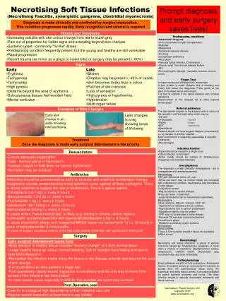
Necrotising Soft Tissue Infections (Necrotising Fasciitis, synergistic gangrene, clostridial myonecrosis). Prompt diagnosis and early surgery saves lives!. Diagnosis is made clinically and confirmed by surgical exploration.

E N D
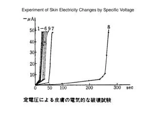
Necrotising Soft Tissue Infections (Necrotising Fasciitis, synergistic gangrene, clostridial myonecrosis) Prompt diagnosis and early surgery saves lives! Diagnosis is made clinically and confirmed by surgical exploration. This condition progresses rapidly. Early recognition and referral is required • History and Symptoms • Spreading cellulitis with skin colour change from red to bluish grey • Pain out of proportion for visible signs and extending beyond skin changes • Systemic upset -commonly "flu like" illness • Predisposing condition frequently present but the young and healthy are still vulnerable • Rapid progression • Recent trauma (as minor as a graze or insect bite) or surgery may be present (~80%) • Predisposing conditions • Intravenous drug use • Immunosupression/organ transplantation • Diabetes • Malignancy • Peripheral vascular disease • Smoking • Complement deficiency • Alcoholism • Varicella Zoster Infection (Chickenpox) • Chronic renal, liver & heart disease Failure • HIV • Pre-existing skin disease - psoriasis, eczema, chronic ulcers • Finger Test • A suspected area is infiltrated with anaesthetic • A 2cm incision is made. Relative lack of bleeding and murky fluid favour the diagnosis. Probe gently at the level of the deep fascia with a finger. • The test is positive if the tissue dissects with minimal resistance • Discolouration of the deepest fat is often present (brown/grey) • Referral pathways • The appropriate surgeon is the surgeon able to carry out the operation with the least delay which may be: • General • Orthopaedic Insert • Plastic page • ENT numbers • Maxillofacial • Patients should not have surgery delayed unnecessarily i.e. by transfer to another hospital. • Early involvement of supporting specialities is required • Intensivist • Microbiologist • Infection Control • Patient should be nursed in a single room • Inform infection control team • Public health should be notified of Streptococcus Pyogenes and clostridial infections • Investigations • The diagnosis is made clinically. Investigations aid in management and assessing severity. • Haematology • FBC with differential white cell count • -White cell count may be normal initially but increases with a worsening condition. Neutropenia may be present in later stages • Coagulation screen • -May be altered in severe disease • Group and save / crossmatch • -Large transfusions can be required peri-operatively • Biochemistry • U&Es, Glucose, Albumin, Calcium, CRP, CK • -Hyponatremia indicates a worse condition • -Hypocalcaemia due to calcium precipitation • -Hypoalbuminaemia may be present • -CRP may not be elevated in early disease • -Elevated CK indicates muscle involvement • Arterial blood gases • -Metabolic acidosis as sepsis worsens • Microbiology • Blood cultures • Tissue & fluid samples (swabs if tissue not available) • MRSA screen • Bacteriology • Necrotising soft tissue infections: a group of serious infections caused by Streptococcus pyogenes, or more often a mixture of organisms: Staphylococcus aureus, Streptococci, ‘coliform bacilli’, Bacteroides spp, Clostridium spp and other anaerobes. • Pathophysiology • Exact pathways are still not fully understood. Antecedent trauma (if present) allows an entry portal for bacteria to spread from the subcutaneous tissue along the superficial and deep fascial planes. A process facilitated by potent bacterial enzymes and toxins. The deep infection results in vascular thrombosis, ischaemia and tissue necrosis. Signs • Early • Erythema • Tachypnoea • Tachycardia • High pyrexia • Oedema beyond the area of erythema • Subcutaneous tissues feel wooden-hard • Mental confusion • Late • Blisters • Crepitus may be present (~40% of cases) • Skin becomes dusky blue in colour • Patches of skin necrosis • Loss of sensation • High pyrexia or hypothermia • Hypotension • Multi-organ failure Examples of Skin Changes Early skin change in an axilla showing mild erythema. Later changes in a leg showing areas of blistering Treatment Once the diagnosis is made early surgical debridement is the priority Resuscitation • Ensure adequate oxygenation • Fluid - Normal saline or Hartmann's • Inotropic support if fluid does not correct hypotension • Ventilation may be required Antibiotics • Antibiotics should be commenced as early as possible with empirical combination therapy designed to provide comprehensive broad spectrum cover against all likely pathogens. There is strong evidence to support the use of clindamycin. This is a typical regime: • Clindamycin 0.6-1.2g i.v. every 6 hours • Benzylpenicillin 1.2-2.4g i.v. every 4 hours • Flucloxacillin 1-2g i.v. every 6 hours • Gentamicin 120-160mg i.v. every 12 hours • Metronidazole 500mg i.v. every 8 hours • In cases where Pseudomonas spp. is likely (e.g. arising in chronic ulcers) replace flucloxacillin and benzylpenicillin with piperacillin/tazobactam 4.5g i.v. 8 hourly. • In cases of penicillin allergy or in suspected MRSA cases use vancomycin 1g i.v. 12 hourly in place of benzylpenecillin & flucloxacillin • (In cases of suspect cutaneous anthrax which has some clinical similarities add ciprofloxacin 400mg bd) Surgery • Early surgical debridement saves lives • Wide excision to healthy tissue consider "excision margin" of 2.5cm normal tissue • Judge healthy tissue by colour, normal bleeding, lack of wooden-hard feeling and lack of easy blunt dissection. • Remember the infection tracks along the fascia so the disease extends well beyond the area of skin change • If in doubt about an area perform a finger test • Post-operatively regular wound inspection is mandatory and the only way to know if the spread of the infection has been halted • In more severe cases especially in IVDUs the muscles are commonly involved Post Operative care Canniesburn Plastic Surgery Unit Copyright 2005 www.canniesburn.org • Care for in a surgical high dependency unit or intensive care unit • Regular wound inspection at least twice a day initially