Classical Genetics Principles
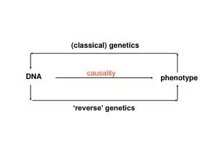
Classical Genetics Principles. BIT 220 Chapters 4-7, part of 9. End of chapter 3. P. 63-end - Mendel and Human Genetics Pedigree analysis - diagrams that show the relationships - Figure 3.13/3.15

Classical Genetics Principles
E N D
Presentation Transcript
Classical Genetics Principles BIT 220 Chapters 4-7, part of 9
End of chapter 3 • P. 63-end - Mendel and Human Genetics • Pedigree analysis - diagrams that show the relationships - Figure 3.13/3.15 • Pedigree conventions- squares for male, circles for female; different colors with individual who express the trait
Chapter 4 - Extensions of Mendelism • Figure 4.1 - Incomplete or Partially dominant (note in this figure new designation) - can also be called semidominant • different amounts of gene product produced
Multiple Alleles • - antigen reacts with serum blood factors • Table 4.1 (p. 75)- ABO blood typing system • Alleles (alternate forms of the same gene) listed as IA, IB, or ii • can have A, B, AB, and O • A and B said to be codominant - both expressed.
Allelic Series • Figure 4.4 - coat colors in animals • many alleles involved- by making heterozygotes, dominance relationships can be determined • See Figure 4.3 for color descriptions • Wild-type allele completely dominant over all other alleles • c+ > cch > ch > c : shows effect on coat color
Testing Mutations for Allelism • Mutant allele - created when existing allele changes to a new genetic state • always different genetic composition; may be different phenotype • Can test mutation if recessive - Figure 4.5 - much like the complementation test - applies more to higher organisms (complementation in bacteria)
Allelism • Another example Figure 4.6 - Drosophila • cinnabar & scarlet (eye color) • cross homozygous mutant strains with each other • shows allelism many more than one eye color gene involved
Types of Mutations • Visible- change appearance of organism usually recessive • Sterile - limit reproduction - recessive or dominant, affect males and females • Lethal - kill organism - interfere with a vital function; Dominant disappears after one generation; recessives show up normal frequency - Figure 4.7 - dominant visible and recessive lethal
Why Dominant or Recessive? • Recessive: loss-of-function - affect only homozygotes • Dominant - affect both heterozygotes and homozygotes - dominant- negative or gain-of-function • Explained in Figure 4.9
From Genotype to Phenotype • Environmental effects - diet and PKU - babies tested early so low phenylalanine diet = normal development • Pattern baldness - testosterone levels equate with more hair loss (why worse in men than women)
Penetrance and Expressivity • Incomplete penetrance - mutation does not show appropriate phenotype even though genetic mutation is present • Example is Figure4.11 • Expressivity - variable phenotype among individual with same genotype - Figure 14.12 - Dominant Lobe mutation in Drosophila
Gene Interactions • Traits can be influenced by more than one gene • Punnett square Figures 4.14 and Figure 4.13 - comb shapes in chickens - crosses between rose and pea produce another type, called walnut - 2 independently assorting genes, each with 2 alleles
Epistasis • Two or more genes influence a trait • An allele of one has overriding effect on phenotype - it is epistatic to other genes involved in the trait (Greek work to “stand above” • Conceals the presence of another mutation in the same gene • Example Figure 14.15 - Lathyrus odoratus (sweet pea)
Pleiotropy • Gene affects many phenotypes • Greek “to take many turns” • PKU - mutation also interferes with melanin synthesis (color pigment) • PKU sufferers have light hair • Blood and urine of PKU sufferers additional compounds absent in non-PKU individuals
Chapter 5 - Inheritance of Complex Traits • Not covering much of this chapter • Know pages 90-92 • Quantitative traits - accumulation of many genes that influence a trait - example is Figure 5.1 - 3 genes with independent assortment and incomplete dominance accounts for kernel color variation • Table 5.4, page 105
Effects of Inbreeding • Pages 105 - 107 • Consanguineous (Latin for “same blood”) mating - mating between relatives • Pedigree Figure 5.9, page 106 - all homozygous albino individuals from consanguineous matings • Amish, Mormon, French Canadians, Royalty
Chapter 6 Chromosomal Basis of Mendelism • Chromosomes - light and dark regions when stained - euchromatin (light), heterochromatin (dark) • Number - Table 6.1, varies among species • Number: • haploid - n (usually 1/2 the full complement) • diploid - 2n • tetraploid - 4n, etc.
Sex Chromosomes • X - females - XX • Y - male - XY • Sex chromosomes and autosomes
Chromosomes • Arrays of genes - Figure 6.5 • Morgan and colleagues - Drosophila • Locus (loci) - where a gene is located on a chromosome • produced map like one in figure for genetic loci • Chromosome theory (heredity) proved by non-disjunction - Figure 6.6
Mendel’s Laws • Segregation - Figure 6.7 - based on separation of chromosomes during anaphase (first meiotic division) • Independent Assortment - Figure 6.8 -random alignment of different pairs of chromosomes at metaphase (genes on same chromosome linked, so don’t assort independently
Sex-Linkage in Humans • Male needs only one recessive gene to express phenotype; female needs 2 • Hemophilia, color blindness, examples of sex-linkage • Figure 6.9 - shows pedigree of Czar Nicholas II - only males have the disease; females are carriers • Figure 6.10 shows color blindness
Fragile X syndrome • Causes mental retardation - appears to follow an X-linked inheritance • abnormality at tip of X chromosome- looks like tip is ready to detach (chromosome doesn’t really brake) • 1/2000 incidence; expansion of repeats • incomplete penetrance - Figure 6.11 • No treatment currently (diagnostic test)
Sex Determination in Humans • Absence of Y chromosome = female • XO and XXX are females • XXY are males • product of SRY (sex-determining region Y) gene - testis determining factor (TDF) - found on short arm of Y chromosome • Figure 6.12
Sex Determination in Drosophila • Y chromosome no role in sex determination • Sex determined by ratio of X chromosomes to autosomes • Flies have XX or XY; 3 pairs of autosomes (3 pairs considered 1 set) A considered one haploid set – Table 6.2, page 128 • When X’s to A’s is >/= 1, fly is female • X to A is </= 0.5, fly is male • between 0.5 and 1 is both sexes
Dosage Compensation of X-Linked Genes • In Drosophila, increase of activity of X-chromosome genes in males (hyperactivation) • In placental mammals, X-linked genes are inactivated on one of the X chromosomes (inactivation); Reactivated during oogenesis (Figure 6.18) • Inactivated X does not look or behave like a normal X – Barr body (Murray Barr)
Chapter 7 Variation in Chromosome Number & Structure • Cytological staining – stains to reveal chromosome banding • Cytogenesis – analysis of stained chromosomes • Compound with insertional power – intercalating agent (Ethidium bromide in DNA, Quinacrine, chromosomes
Banding patterns • Stained chromosomes light and dark bands • Q banding - Quinacrine • G banding – Giemsa • R banding – R banding – opposite of G • C banding – stains region around centromere
Human Karyotype • See Figure 7.4 and 7.5 • XY and 22 autosomes • Can see gross abnormalities this way – • Down syndrome (extra chromosome 21) • Fragile X syndrome • Cri-du-chat – deletion in short arm of Chromosome 5 (cry of the cat) – Figure 7.16 • Can look at rearrangements
Ploidy • Greek word “fold” • Changes in number of chromosome in cells • Euploid – complete, normal set of chromosomes (humans diploid) • Polyploid – organisms that are not diploid normally (triploids (3n) , tetraploid (4n), etc. – more common in plants than animals
Aneuploidy • A numerical change in part of the genome – change in dosage of a single chromosome • E.g., Down syndrome, Trisomy 21 (3 copies of chromosome 21) – this called a trisomy • Other examples: • 47, XXY (Klinefelter’s) • 47, X (Turner’s) – this one a monosomy
Other chromosome abnormalities • Deletion - Cri-du-chat –short arm chromo 5 • Duplication – extra chromosome segment • Inversions – Figure 7.19 –chromosome segments are flipped around • Translocations – segment detached and reattaches to another chromosome (Figure 7.21) • Reciprocal translocations – Figure 7.22
Chapter 9 – pages 188-196Linkage analysis in Humans • Knowledge of genes and their chromosomal location • Knowledge of relationships from one gene to another • Genes on X chromosome easiest to find- see inheritance pattern more clearly • Sidelight – linkage between hemophilia and color blindness (page 189)