Download
1 / 49
550 likes | 1.49k Views
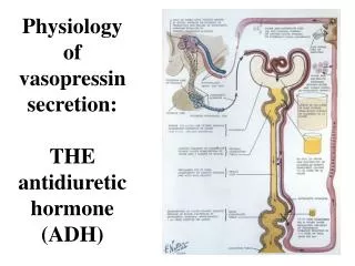
Antidiuretic hormone and the mineralcorticoids . Antidiuretic Hormone: ADH. ADH is also known as arginine vasopressin (AVP = ADH) because of its vasopressive activity, but its major effect is on the kidney in preventing water loss. . Synthesis of ADH.

E N D
Antidiuretic Hormone: ADH • ADH is also known as arginine vasopressin (AVP = ADH) because of its vasopressive activity, but its major effect is on the kidney in preventing water loss.
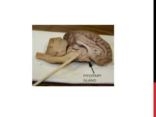
Synthesis of ADH • It is synthesized as pre-prohormones and processed into a nonapeptide (nine amino acids). • Six of the amino acids form a ring structure, joined by disulfide bonds. • It is very similar in structure to oxytocin, differing only in amino acid #3 and #8. • ADH synthesized in the cell bodies of hypothalamic neurons in the supraoptic nucleus • ADH is stored in the neurohypophysis (posterior pituitary)—forms the most readily released ADH pool
Synthesis of ADH • Mechanical disruption or the neurohypohyseal tract by trauma, tumor, or surgery temporarily causes ADH deficiency. • ADH will be restored after regeneration of the axons (about 2 weeks). • But if disruption happens at a high enough level, the cell bodies die in the hypothalamus resulting in permanent ADH deficiency
Secretion of ADH • The biological action of ADH is to conserve body water and regulate tonicity of body fluids. • It is primarily regulated by osmotic and volume stimuli. • Water deprivation increases osmolality of plasma which activates hypothalmic osmoreceptors to stimulate ADH release.
Secretion of ADH • Conversely, water ingestion suppresses osmoreceptor firing and consequently shuts off ADH release. • ADH is initially suppressed by reflex neural stimulation shortly after water is swallowed. • Plasma ADH then declines further after water is absorbed and osmolality falls
Pathway by which ADH secretion is lowered and water exrection raised when excess water is ingested
Secretion of ADH • If plasma osmolality is directly increased by administration of solutes, only those solutes that do not freely or rapidly penetrate cell membranes, such as sodium, cause ADH release. • Conversely, substances that enter cells rapidly, such as urea, do not change osmotic equilibrium and thus do not stimulate ADH release. • ADH secretion is exquisitely sensitive to changes in osmolality. • Changes of 1-2% result in increased ADH secretion.
Secretion of ADH • ADH is stimulated by a decrease in blood volume, cardiac output, or blood pressure. • Hemorrhage is a potent stimulus of ADH release. • Activities, which reduce blood pressure, increase ADH secretion. • Conversely, activities or agents that increase blood pressure, suppresses ADH secretion.
Pathway by which ADH secretion and tubular permeability to water is increased when plasma volume decreases
Secretion of ADH • Hypovolemia is perceived by “pressure receptors” -- carotid and aortic baroreceptors, stretch receptors in left atrium and pulmonary veins, and juxtaglerular apparatus of the kidney. • Normally, pressure receptors tonically inhibit ADH release. • Decrease in blood pressure induces ADH secretion by reducing input from pressure receptors. • The reduced neural input to baroreceptors relieves the source of tonic inhibition on hypothalamic cells that secrete ADH.
Hypothalamus, posterior pituitary and ADH secretion– connection with baroreceptors
Secretion of ADH • Hypovolemia also stimulates the generation of renin and angiotensin directly within the brain. • This local angiotensin II enhances ADH release in addition to stimulating thirst. • Volume regulation is also reinforced by atrial naturetic peptide (ANP). • When circulating volume is increased, ANP is released by cardiac myocytes, this ANP along with the ANP produced locally in the brain, acts to inhibit ADH release.
Secretion of ADH • The two major stimuli of ADH secretion interact. • Changes in volume reinforce osmolar changes. • Hypovolemia sensitizes the ADH response to hyperosmolarity.
Osmotic and hemodynamic control of ADH secretion– changes in osmolality; changes in blood volume or pressure; interactions between osmolar and volume/pressure stimuli on ADH secretion.
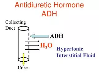
Actions of ADH • The major action of ADH is on renal cells that are responsible for reabsorbing free (osmotically unencumbered) water from the glomerular filtrate. • ADH responsive cells line the distal convoluted tubules and collecting ducts of the renal medulla. • ADH increases the permeability of these cells to water. • The increase in membrane permeability to water permits back diffusion of water along an osmotic gradient. • ADH significantly reduces free-water clearance by the kidney
Actions of ADH • ADH action in the kidney is mediated by its binding to V2 receptors, coupled to adenylate cyclase and cAMP production. • cAMP activates protein kinase A which prompts the insertion of water channels into the apical membrane of the cell. • When ADH is removed, the water channels withdraw from the membrane and the apical surface of the cell becomes impermeable to water once again. .
Actions of ADH • This mechanism of shuttling water channels into and out of the apical membrane provides a very rapid means to control water permeability • The basolateral membrane of the ductal cells are freely permeable to water, so any water that enters via the apical membrane exits the cell across the basolateral membrane, resulting in the net absorption of water from the tubule lumen into the peritubular blood.
Actions of ADH • Water deprivation stimulates ADH secretion, decreases free-water clearance, and enhances water conservation. ADH and water form a negative feedback loop.
Actions of ADH • ADH deficiency is caused by destruction or dysfunction of the supraoptic and parventricular nuclei of the hypothalamus. Inability to produce concentrated urine is a hallmark of ADH deficiency and is referred to as diabetes insipidus. • ADH also acts on the anterior pituitary to stimulate the secretion of ACTH.
Pathways by which sodium and water excretion are decreased in response to severe sweating.
Aldosterone and the mineralocorticoids • The mineralocorticoid, aldosterone is vital to maintaining sodium and potassium balance and extracellular fluid volume. • Aldosterone is an adrenal corticosteroid, synthesized and secreted by the adrenal cortex.
Cross section through the adrenal gland– cortex and medulla salt sugar sex
Aldosterone • The adrenal cortex is composed of three major zones, differentiated by the histological appearance and type of corticosteroid they produce. • The outermost is the zona glomerulosa, is very thin and consists of small cells with elongated mitochondria.
Adrenal zones • The middle zona fasiculata is the widest zone and consists of columnar cells that are highly vacuolated with numerous lipid droplets. • These lipid droplets are composed of cholesterol esters the substrate for adrenal steroid hormone biosynthesis.
Adrenal zones • The innermost zona reticularis contains fewer lipid droplets than fasiculata cells, but have similar mitochondria. • ACTH has trophic effects on the zona fasiculata and reticularis.
Aldosterone synthesis • Aldosterone is synthesized and secreted by the zona glomerulosa . • The synthesis of aldosterone from cholesterol to corticosterone is identical to the synthesis of glucocorticoids in the zona fasiculata. • The C18 methyl group of corticosterone is hydroxylated and converted to an aldehyde yielding aldosterone.
Aldosterone’s function • The principal function of aldosterone is to sustain extracellular fluid volume by conserving body sodium. • Aldosterone is largely secreted in response to signals that arise from the kidney when a reduction in circulating fluid volume is sensed. • When body sodium is depleted, the fall in extracellular fluid and plasma volume decreases renal arterial blood flow and pressure.
Regulation of aldosterone secretion: Activation of renin-angiotensin system in response to hypovolemia is predominant stimulus for aldosterone synthesis.
Aldosterone and renin-angII • The justaglomerular cells of the kidney respond to this change by secreting renin. Renin acts on angiotensinogen (which is secreted by the liver) to form angiotensin I which is further cleaved by angiotensin converting enzyme (which is secreted by the lungs) to angiotensin II.
Aldosterone • Angiotensin II acts on the zona glomerulosa to stimulate aldosterone synthesis. • Angiotensin II acts via increased intracellular cAMP to stimulate aldosterone synthesis.
Aldosterone • ANP reinforces the effects of the renin-angiotensin system on aldosterone secretion. • In response to volume expansion, artrial myocytes secrete ANP which binds to receptors in the zona glomerulosa to inhibit aldosterone synthesis. • ANP acts via increased intracellular cGMP which opposes cAMP and inhibits aldosterone synthesis. • ANP also reduces aldosterone indirectly by inhibiting renin release.
Aldosterone clears potassium • Aldosterone facilitates the clearance of potassium from the extracellular fluid, and potassium stimulates aldosterone synthesis—thus providing a feedback control mechanism to control potassium levels. • Converesly, potassium depletion lowers aldosterone secretion. • Potassium stimulates aldosterone synthesis by depolarizing zona glomerulosa cell membranes to stimulate aldosterone synthesis.
Aldosterone synthesis • Voltage-sensitive calcium channels open and the intracellular calcium concentration increases—activating calcium calmodulin kinase. • Calcium channel blockers inhibit aldosterone synthesis.
Aldosterone synthesis • ACTH also stimulates aldosterone synthesis. • However the ACTH stimulation is more transient than the other stimuli and is diminished within several days. • ACTH provides a tonic control of aldosterone synthesis. • In the absence of ACTH, sodium depletion still activates renin-angiotensin system to stimulate aldosterone synthesis. • Aldosterone levels fluctuate diurnally—highest concentration being at 8 AM, lowest at 11 PM, in parallel to cortisol rhythms.
Aldosterone action • Aldosterone binds to the mineralocorticoid receptor in target cells and affects transcriptional changes typical of steroid hormone action. • The kidney is the major site of mineralocorticoid activity.
Model for basic renal handling of potassium. Net absorption occurs in the proximal portions of the nephron and net secretion in the more distal portions of the nephron.
Action of aldosterone on the renal tubule. Sodium reabsorption from tubular urine into the tubular cells is stimulated. At the same time, potassium secretion from the tubular cell into urine is increased. Na+/K+-ATPase, and Na+ channels work together to increase volume and pressure, and decrease K+.
Aldosterone action • Aldosterone stimulates the active reaborsption of sodium from the tubular urine back into the nearby capillaries in the distal tubule. • Water is passively reabsorbed with sodium which maintains sodium concentrations at a constant level. • Hence extracellular fluid volume expands in a virtually isotonic fashion
Aldosterone action • Aldosterone stimulates the active secretion of potassium from the tubular cell into the urine. • Most potassium that is excreted daily results from distal tubular secretion. • Hence aldosterone is critical for disposal of daily dietary potassium load at normal plasma potassium concentrations.
Pathway by which an increased potassium intake induces greater potassium excretion mediated by aldosterone
Pathway by which aldosterone sercretion and tubular sodium reabsorption is increased when plasma volume is decreased
Aldosterone action • Increased blood pressure results from excess aldosterone. • Hypertension is an indirect consequence of sodium retention and expansion of extracellular fluid volume.
Cortisol is at 1000 fold higher concentrations than aldosterone
Aldosterone action • Cortisol binds well to the mineralocorticoid receptor and plasma cortisol levels are orders of magnitude higher than aldosterone. • Target tissues for aldosterone are protected from glucocorticoid excess via the action of 11b-hydroxysteroid dehydrogenase, the enzyme that converts cortisol to cortisone, a biological inactive metabolite. • Aldosterone is not a substrate for 11b-HSD and thus only it can bind to its receptor.